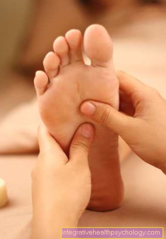
Foot muscles
definition

There are skeletal muscles on the foot as on other parts of the body.
These foot muscles are topographically divided into the muscles of the back of the foot (Dorsum pedis), the sole of the foot (Planta pedis).
Furthermore, a distinction is made between the muscles of the sole of the foot between the big toe and the little toe muscles and the muscles in the middle area. In general, a distinction is made between short and long muscles on the foot. The muscle bellies of the long foot muscles lie on the lower leg and only their tendons pull along the foot. The muscles in the foot enable the foot to move in various degrees and are also involved in stabilizing the foot when walking.
Short foot muscles
The short muscles of the back of the foot are on top of the bone and are also known as the intrinsic muscle group because they lie right in the area of the foot.
One distinguishes one Toe extender, the alone pulls to the big toe. This is called Extensor hallucis brevis muscle and is responsible for the movement, especially the stretching and pulling of the big toe. It runs from the front of the Heel bone (Calcaneus) to the base of the distal phalanx of the big toe.
Besides this Muscles lies the short toe extensors, which in medical terminology as Extensor digitorum brevis muscle referred to as. This also has its origin at the front of the calcaneus and is divided into three muscle bellies. A tendon emerges from each of these muscle bellies, which attaches relatively far forward to the second to fourth toes.
This muscle also does the stretching and that Tightening the toes towards the lower leg. This movement will Dorsiflexion called. It is a flexion in the direction of the back of the foot. The small and therefore fifth toe often does not have a tendon that extends from the muscle. A tendon is very rarely present and in general the anatomy can be very different, so that some tendons can be missing.
Both of these muscles are used by the same nerves, the Fibular nerve excited, whereby the movement can be carried out.
Long muscles of the back of the foot
The longer muscles of the foot are characterized by the fact that they have a very long course and their long tendons over the Back of the foot to the toes run away.
Since they lie outside the foot and also have their origin there, they are called extrinsic muscles and are the counterpart to the intrinsic muscles.
The long toe extensor pulls from the outside of the Knee joint bone oblique to the region of the ankle joint. Eventually one comes out of the relatively small muscle belly long tendon emerged. At about the level of the ankle, the tendon divides into 4 further partial tendons. These tendons now run along the end members of the second to fifth toes.
The muscle becomes like the short muscles of the foot Fibular nerve excited. The innervation of the Muscle leads to extension and flexion in the direction of the lower leg of the toes it is targeting. Here, too, it is a question of dorsiflexion. Since the muscles from the knee to the Ankle joint runs to the toes, it not only influences the movement of the toes, but also moves the ankle.
There it causes the foot to rotate around the longitudinal axis in such a way that the outer edge of the foot is raised while the inner edge is lowered downwards. The heel does not turn with this. This type of movement is also found in the wrist and elbow joint and is called the Pronation designated. Pronation has that on the foot Synonym Eversion.
Similar to the short muscles on the foot, the long muscles on the foot have a separate muscle that performs the same movement on the big toe. This is the long big toe extensor (Extensor hallucis longus muscle). Its origin lies directly next to the plug for the remaining toes and also extends in its vicinity over the ankle joint to the end joint of the big toe. These muscles result in the same movement as the other muscles on the back of the foot. It also supports pronation in the ankle.
Muscles of the sole of the foot
At the Sole of the foot a distinction is made between other muscle groups.
The muscles lying here are responsible for flexing and pulling the toes. With the diffraction one further distinguishes between one long and short flexors for the big toe and a flexor for the remaining adjacent toes.
Furthermore there are the muscles in the Big toe area, Little toe area and such muscles that between the toes lie.
Big toe muscles
The muscles in this area include the Spreader the big toe, the Abductor hallucis muscle.
This muscle arises from the front side of the heel on the lower surface and pulls to the Sesame bone metatarsal and to the base of the metatarsophalangeal joint of the big toe. These muscles spreads the toe outward away and also contributes to one slight flexion of the big toe.
The opposite movement is caused by the Donners the big toe allows. With this muscle the short flexor of the big toe is also partly fused, the Flexor hallucis brevis muscle. This has its origin in a metatarsal bone. In its course it divides into two muscle tummies, one of which lies further inside, the other further outside. The two muscle bellies divide into two again Partial tendons one of which also attaches to the sesamoid bone and the metacarpophalangeal joint. The flexor muscle is especially important for the flexion of the big toe. This movement is known as planaflexion. The toe is moved away from the lower leg and downwards.
Next to the short flexor muscle there is another long flexor muscle of the big toe. This is not directly counted among the muscles in the big toe area because it originates from the back of the lower leg bone. There it pulls from the outside at an angle to the sole of the foot and finally attaches its tendon to the end joint of the first toe. The muscle has a thicker muscle belly and contributes significantly to the support of the arch of the foot. It is one of the most important muscles around you Flat foot, in which the foot comes to lie incorrectly on the ground, to counteract this.
Middle foot muscles
Between the small and the big toe there are other muscles that partly support the functions of other muscles and partly take on other important tasks to stabilize the arch of the foot.
These include the lumbicral muscles. These four small muscles are located on the inward-facing side of the tendons of the long toe flexor. You support the flexing movement of the toes and at the same time pull the toes towards each other.
The latter movement will also Adduction called. They also increase the stiffness of the arch of the foot and thus contribute to it stability of the entire foot. Individual anatomical differences occur especially with these small muscles. This means that it can be used in both diminished as well as in increased number occurrence.
Another muscle that is very central to the sole of the foot is the Sole quadrilateral muscle (M. quadratus plantae). This muscle is also connected to the lateral outer edge of the long toe flexor and supports its function. It also helps to strengthen the arch of the foot. Furthermore there is small muscles between the individual toes, which act as interbone muscles (Interossei muscles) are designated.
They spread to the Sole of the foot and Back of the foot down. The muscles executed on the sole of the foot cause a attraction of third to fifth toes towards the second toe. They also contribute to a slight flexion in the metatarsophalangeal joints of the toes.
The small muscles that are executed on the back of the foot tend to perform a splaying motion of the toes. The short flexor of the toes, the M. flexor digitorum brevis. A small muscle that pulls from the lower surface of the calcaneus to the middle portion of the bone of the second through fourth toes. The muscle causes the toes to flex. In addition to the short flexor muscle of the toes, there is also a larger and longer one. This is not on the foot itself, but belongs to the rear muscle group on the lower leg. This muscle pulls from the posterior surface of the tibia to the terminal limbs of the second to fifth toes.
In addition to a bending movement, it also helps to rotate the foot around the longitudinal axis. In such a way that the inner foot edge is raised while the outer foot edge is lowered. This movement is known as supination and is the opposite of pronation.
Little toe muscles
There are also in the area of the little toe own musclesused to move the little toe serve. There is also the Little toe opposingthat in medical terminology too M. opponens digiti minimi is called.
After arousal by the appropriate nerve this muscle performs a coherent movement of tightening and flexing the little toe. This movement is called Oppose designated. He also strengthens the arch of the foot. Individual differences can occur in this muscle, so that it is sometimes even absent entirely. The flexor of the little toe, the M. flexor digiti minimi. This flexes the toe in the direction of the sole of the foot.
Often another muscle is fused with this muscle. This is the Spreader of the little toe, the M. abductor digiti minimi. It is the largest and longest muscle in this region and extends from part of the calcaneus and its lower surface to the wrist of the fifth toe. It essentially forms the outer and thus the lateral edge of the foot. This muscle also performs a plantar flexion of the little toe. In addition supports he arches the foot. Although one can deduce from his name, he only performs a splaying movement to a limited extent.


.jpg)