Ear canal
General
The term "ear canal" refers to two different anatomical structures. On the one hand, it describes the "internal auditory canal" (Meatus acousticus internus), on the other hand the "external auditory canal" (Meatus acusicus externus). Colloquially, however, the latter is usually meant.
The external ear canal
construction
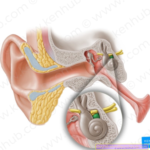
The outer auditory canal as part of the outer ear is approximately 2 - 2.5 cm long in humans and extends from the auricle to the eardrum, which separates it from the so-called middle ear. In the outer third of its course, its wall is formed by cartilage, while the remaining two thirds are bony and part of the temporal bone.
It is completely lined by skin, which ultimately merges into the eardrum. In addition to the skin, bristle hairs protect the outer third (Tragi) our ear canal from foreign bodies. Special sebum glands, the ball glands, together with the secretion of the sebum glands and flaked skin flakes form the ear wax (Cerumen). This also has a protective function by being antibacterial and antifungal and thus counteracting infections.

function
In addition to its function of conveying the sound bundled by the auricle to the eardrum, the external auditory canal is also able to amplify the sound through its own resonance. This happens mainly in the frequency range of 2000 - 4000 Hz, which is why we are more sensitive to these frequencies than to others. So it is not surprising that part of our main language range (the frequency in which we speak, approx. 500 - 3000 Hz) is in this range.
Diseases
Ear canal diseases are often caused by the Using cotton swabs to clean it up. One of the most common diseases caused by this is that Ear canal furuncles. this is a Inflammation of the hair follicles of the ear canal bacteria and is having pain from pressure on that ear or characterized by chewing. It is handled by alcohol-soaked strips and ointments containing antibioticswhich are inserted into the ear.
In the course of the Inserting a cotton swab or of an otoscope by the doctor it may in some people too Throat irritation come. This is because the external ear canal is covered by a branch of the person Innervated nerves is ours too Larynx sensitively innervated and is in no way questionable.
The inner ear canal
In contrast to the external ear canal is the inner ear canal Part of the inner ear and runs in the petrous bone. He serves both Facial nerve (VII. Cranial nerve), Vestibulocochlear nerve (VIII. Cranial nerve), as well Blood vessels as a passage into the posterior fossa. Guide said nerves acoustic stimuli to the brain, or signals from the brain to the facial muscles.


.jpg)