Low-dose CT
definition
With the help of a CT, ionizing radiation is used to obtain high-resolution images of the body. In a low-dose CT, a particularly low radiation dose is used compared to a normal CT. This reduces the risky radiation dose for the patient. The low-dose CT is used, among other things, to detect stones in the kidney. In this case one speaks of a stone CT.
Read more on the subject at: Computed Tomography

Indications
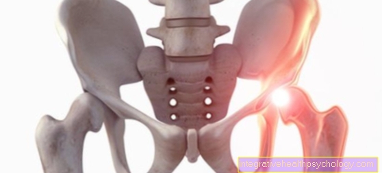
Low-dose CT is used when there is already good contrast. This means that the structures to be examined can be easily distinguished from one another. The stone CT is especially used to detect kidney stones (Urolithiasis) used. Ultrasound is an alternative to this. Its results are, however, inferior to those of the stone CT.
Read more on the subject at: Kidney stones
In patients with an increased risk of lung cancer, such as Long-term heavy smokers, a low-dose CT can be used as screening for early detection. A CT scan of the lungs can detect possible cancer earlier than an X-ray examination. The introduction of an official screening for early lung cancer detection is very controversial. The low-dose CT can also be used if the skeleton is to be examined.
Read more on the subject at: Diagnosis of lung cancer
preparation
If there is a need for a low-dose CT scan, the patient must first be informed about the examination by a doctor. The patient then has to give his consent to the examination by means of a signature. He also has the opportunity to ask questions. Shortly before the examination, objects such as jewelry, glasses, hearing aids must be put down. In addition, an artificial bit must be removed if one is worn. If a contrast agent is given, venous access must also be established.
procedure
The process is similar to that of a normal CT examination. Before the CT examination, all jewelry etc. should be taken off, as these can lead to other structures in the area no longer being displayed correctly. A low-dose CT is performed with a completely normal CT machine on which the settings are adjusted. During the CT examination, the patient lies down. If possible, it should not move, as this affects the image quality. During a CT examination of the lungs, the patient has to hold his breath for a few seconds. While the image is being taken, other people have to leave the room due to the radiation exposure. If the CT image is taken with contrast agent, contrast agent is given through a venous access during the examination.
evaluation
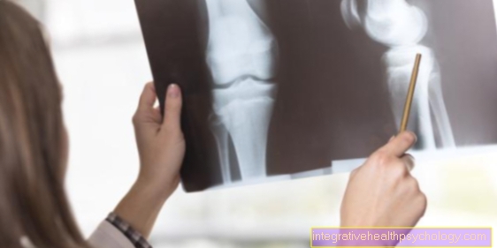
The evaluation of the CT image is carried out by a radiologist. The attending physician receives the result. CT images, including those from a low-dose CT, have a significantly better resolution than normal X-ray images. Therefore, significantly more can be recognized and diagnosed on them. However, the images are not 100% clear in every case either. In the CT image of a lung, it is often not possible to say with certainty whether it is lung cancer or not. Because there are other causes that show up similarly on a lung CT. A questionable lung cancer can then be diagnosed or ruled out through further examinations
Risks
In a CT, radiation is used, whereby on the one hand good examination results are achieved, on the other hand there are risks from the ionizing radiation used. Because radiation has been shown to cause cancer. However, the risk posed by the radiation does not lie in the single but in the repeated use of a CT. One speaks here of the cumulative radiation dose - i.e. the amount of radiation that is accumulated through multiple CT examinations. With increasing radiation exposure, the likelihood that various cancers could develop as a long-term consequence increases. Children are more sensitive to radiation and its risks.
With a low-dose CT, the radiation used is lower. The risk of a single examination is low. In addition, CT examinations achieve very good results, which are often decisive for therapy or life-saving. Any CT exam should weigh how important it is. The principle should be taken into account as little as possible, as often as necessary. The additional administration of contrast media during a CT examination creates additional risks. An allergic reaction can occur. The contrast agent can also damage the kidneys.
How is the radiation exposure?
The radiation exposure depends on which area of the body is examined with the CT. A CT scan of the chest produces 1-10 millisieverts of radiation. An X-ray would only produce 0.01 to 0.1 millisievert. In the case of a low-dose CT, the radiation involved should be below 2 millisievert. Compared to standard CT, up to 60% of the radiation dose can be saved. But even in everyday life you are always exposed to radiation. The radiation with which one is exposed to natural radiation in one year averages 2.1 millisieverts.
Read more on the subject at: Radiation exposure in computed tomography
Duration
A CT scan itself does not take long. Depending on the areas from which the low-dose CT is to be performed, the examination itself is usually completed within a few minutes. However, there are often longer waiting times before the examination. The actual creation of the image, i.e. the scanning of the body with the help of the radiation, is done within fractions of a second. If an additional contrast agent has to be administered, the CT takes a little longer. However, the evaluation of the CT image must then take place, which takes longer than the actual recording of the image. A radiologist can do this in minutes. How long the entire examination actually takes from preparation to result depends on the various waiting times and is different in every practice or clinic.
costs
If there is a medical indication, the health insurance company will cover the costs of the low-dose CT. The cost of the examination varies depending on which area of the body is examined. However, if a long-term smoker without symptoms would like to have a CT scan of his lungs to rule out lung cancer, there is no medical indication for the health insurance company, as there are no underlying diseases and no complaints. In this case, the patient very often has to bear the costs himself. In many cases, however, private health insurance companies will still cover the costs.
Do i need contrast media?
Whether a contrast agent is needed depends on the medical question. The administration of contrast media is important for diagnosing some diseases. If the administration of contrast medium makes sense for your medical question, your attending physician will explain this to you. As a rule, contrast agent is not necessary to identify kidney stones. Contrast media is required, however, when the lower urinary tract is to be displayed, e.g. plan a procedure for treatment. If vessels are to be displayed in a low-dose CT examination, i.e. a so-called CT angiography is performed, contrast agent must be given. Without this one cannot adequately assess the vessels.
Further informationFor more information, see:
- Computed Tomography
- CT of the lungs
- CT of the chest
- CT abdomen
- Radiation exposure in computed tomography
You can find an overview of all topics in the field of diagnostics under: Diagnostics A-Z