Syndesmoser crack
definition

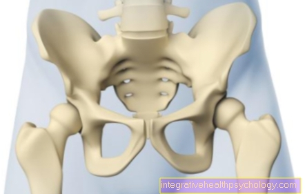
As syndesmosis (Membrana interossea) is the term used to describe the connective tissue membrane that connects the fibula and shinbone and is therefore required to stabilize the ankle joint.In the lower part near the ankle joint, the syndesmosis guarantees this stability in cooperation with the outer and inner ligaments.
At Twisting or compression injuries of the ankle joint, the syndesmosis (syndesmosis rupture) or part of it can occur. Especially one excessive external rotation of the talus (Talus) holds the Risk of syndesmosis injury.
This can also occur when no bony structures affected so that the exclusion of a fracture in the area of the ankle is not sufficient to rule out a syndesmosis tear.
Emergence
The syndesmosis tear is one classic sports injury and joins frequently sports on in which accidents with severe twisting of the ankle are likely. These include, for example To ski in the same way as motion-intensive ball sports.
Appointment with ?

I would be happy to advise you!
Who am I?
My name is I am a specialist in orthopedics and the founder of .
Various television programs and print media report regularly about my work. On HR television you can see me every 6 weeks live on "Hallo Hessen".
But now enough is indicated ;-)
Athletes (joggers, soccer players, etc.) are particularly often affected by diseases of the foot. In some cases, the cause of the foot discomfort cannot be identified at first.
Therefore, the treatment of the foot (e.g. Achilles tendonitis, heel spurs, etc.) requires a lot of experience.
I focus on a wide variety of foot diseases.
The aim of every treatment is treatment without surgery with a complete recovery of performance.
Which therapy achieves the best results in the long term can only be determined after looking at all of the information (Examination, X-ray, ultrasound, MRI, etc.) be assessed.
You can find me in:
- - your orthopedic surgeon
14
Directly to the online appointment arrangement
Unfortunately, it is currently only possible to make an appointment with private health insurers. I hope for your understanding!
Further information about myself can be found at
diagnosis
A syndesmosis rupture sometimes causes strong pain when stressing the ankle, one Full load of the affected leg often impossible power. The joint also swells strongly in most cases and has a Tenderness as well as a painful external rotation on.
Since the radiological Rule out a bone injury Injury to the ligamentous apparatus does not exclude the diagnosis should primarily be made clinically.
The local tenderness and the Behavior in the stress test external rotation of the ankle or compression of the tibia and fibula (syndesmosis compression test) can provide an indication of the extent of the injury.
Conventional radiographs and magnetic resonance imaging (MRIs) are often helpful and can support the diagnosis. Magnetic resonance imaging can also be useful in Planning a possibly necessary operation be.
If a syndesmosis tear is suspected, other injuries such as a torn lateral ligament, ankle fracture and fractures of the tibia or fibula can also be considered and must be ruled out.
therapy
In the acute phase must the affected extremity elevated and cooled become. In addition, the ingestion of Anti-inflammatory drugs (Ibuproipfen, Paracetamol, aspirin) useful for pain therapy.
The primary goal of long-term therapy is that Restoring the stability of the ankle and thus the ability to exercise and exercise. Here is between the strain or the incomplete tearing the syndesmosis and the complete disruption to differentiate. In the case of an incomplete tear, conservative care can be given Immobilization in an orthosis respectively. In the event of a complete rupture of the syndesmosis, combined with severe pain and instability of the upper ankle, the conservative regime can be extended.
The affected limb is attached to a Lower leg walking cast or one removable brace For about six to ten weeks immobilized.
The exercise can be done at lack of tenderness and if external rotation of the calcaneus is possible without pain and should be carried out by a Physiotherapists be accompanied as soon as full resilience exists.
At a gross misalignment of the calcaneus may be a operative therapy necessary. Depending on the procedure, the joint is treated with a ankle-spanning adjusting screw stabilized and the ligamentous apparatus, if necessary, reconstructed with absorbable sutures or a minimally invasive procedure performed using permanent implants that remain in the body. In the first case, metal removal under short-term anesthesia is necessary at a later point in time, before the start of full exercise.
Postoperatively, with the help of forearm crutches, partial load on the injured joint is possible.
Forecast: ability to work
To one to two weeks can do sedentary activities like Employment at the desk and office work to be resumed. When moving around the workplace, the consistent use of walking aids to be observed.
Standing activities must first be avoided. A possible use in the workplace depends on the clinic of the injured extremity. Work that involves greater physical exertion may only be resumed when full exertion is possible and adequate physiotherapeutic therapy has taken place.
Forecast: ability to exercise
To six weeks can the careful, therapeutic training to be resumed. It is important to ensure that the clinic of the joint enables the therapy. Swelling and pain should be reduced as much as possible, as described above.
When carried out consistently and professionally Reloading Restoration of athletic performance can be expected after ten to twelve weeks to the extent that it existed before the injury.
When treating the injured, often young, one has to pay attention to a careful and above all adapted procedure in order to avoid a deterioration of the injury pattern with possible long-term damage.