Ultrasonic
Synonyms in a broader sense
Ultrasound examination, sonography, sonography
definition
Sonography or ultrasound examination is the use of ultrasound waves to examine organic tissue in medicine. A sonogram / ultrasound is an image that is created with the help of sonography.
The investigation works with inaudible sound waves on the echo principle, comparable to the echo sounder in seafaring.

Basics and technology
From a physical point of view, ultrasound describes sound waves above the human hearing range. The human ear can perceive sounds up to approx. 16-18,000 Hz. The ultrasonic range is between 20,000 Hz - 1000 MHz. Bats use ultrasound waves for orientation in the dark. Sounds of even higher frequencies are called hypersonic. Below the sound that can be heard by humans, one speaks of infrasound.
Ultrasonic waves from the sonography device are generated with so-called piezoelectric crystals. Piezoelectric crystals vibrate during Ultrasonic while applying a corresponding alternating voltage and thus emit the ultrasonic waves.
A requirement for the ultrasound examination in medicine is liquid. Air-filled cavities like lung and Intestines cannot be examined and assessed, or only to a limited extent.
In the ultrasound examination, the ultrasound head, which is both transmitter and receiver, sends an ultrasound pulse into the tissue. If this is reflected in the tissue, the impulse comes back and is registered by the receiver. The depth of the reflected tissue can be made over the length of the run over the duration of the transmitted pulse and registration via the receiver.
Procedure

The introduction of the Ultrasound diagnostics in the Orthopedics goes back to Prof. R. Graf 1978. Graf began to sound the child's hip joint in order to be able to recognize hip dysplasias in infancy X-rays do not provide any information because of the missing skeleton. The indication for the use of sonography in the Orthopedics continuously larger (please refer Indications).
The so-called B-mode is generally used for the investigation. Not a single impulse is sent, but a “pulse wall” is used over a line of several centimeters.As a result, the sonic device calculates a layer image of the ultrasound tissue.
In the Orthopedics Depending on the required penetration depth, transducers with frequencies between 5 - 10 MHz for a Ultrasonic used.
Procedure of the investigation
The one with the Ultrasonic The area to be examined is first covered with a gel. The gel is needed because air must be avoided between the tissue and the transducer.
The examination is carried out with light pressure on the tissue. The structures to be examined are scanned in a fan shape in different directions and the joint position is changed. Finally, all structures under movement of joints are assessed.
Regardless of the organ / tissue being scanned, an ultrasound examination always proceeds in the same way: Depending on the structure to be examined, the patient lies down or sits down on an examination couch. The only thing to note here is that the patient should have a Ultrasound of the abdomen (Abdominal ultrasound) be scheduled for this investigation sober appears that the air that would be in the gastrointestinal tract due to previous food intake would interfere with the recorded ultrasound image. First, the doctor applies a gel to the skin that is above the structure to be examined. This gel has a high Water content, which prevents sound from being reflected from air pockets between the surface of the skin and the air. This is the only way to produce a usable image, which is why the examiner must always ensure that there is no air between the gel and the transducer. As soon as the gel layer becomes too thin, the image deteriorates, so that it is sometimes necessary to reapply gel several times during an examination.
The crucial device of the ultrasound examination is the so-called Transducerthat sometimes too probe is called. This is connected via a cable to the actual ultrasound device, on which there is a monitor on which the recorded image can be seen. In addition, this device is operated using several buttons that make it possible, for example, to change the brightness, create a still image or a Color Doppler (see below) over the picture. The probe is responsible both for sending the ultrasound and for receiving it again after it has been reflected.
There are different types of probes. One distinguishes Sector, linear and convex probeswhich are used in different areas due to their different properties. The sector probe has only a small coupling surface, which is useful when you are looking at structures that are difficult to access, such as the heart want to investigate. When using sector probes, the typical fan-shaped ultrasound image is created on the screen. A disadvantage of these probes, however, is that poor image resolution near the transducer.
The Linear probes have a large contact area and parallel sound propagation, which is why the resulting image is rectangular. This gives them good resolution and is particularly suitable for superficial tissue like the thyroid to investigate.
The Convex probe is practically a combination of sector and linear probe. In addition, there are some special probes, for example the TEE probethat is swallowed that Vaginal probe, the Rectal probe and the Intravascular ultrasound (IVUS), in which thin probes can be inserted directly into the vessels. In any case, the probe is usually placed on the gel previously applied to the body. The desired structure can then be targeted by moving the probe back and forth or angling it. The transducer now sends out short, directed sound wave pulses. These waves are reflected or scattered more or less strongly by the successive different layers of tissue. This phenomenon is known as Echogenicity. The transducer now serves not only as a sound transmitter but also as a receiver. So it picks up the reflected rays again. A reconstruction of the reflecting object can thus take place from the transit time of the reflected signals. The reflected sound waves are converted into electrical impulses, then amplified and then displayed on the screen on the ultrasound device.
A low echogenicity demonstrate liquids (for example blood or urine), these are shown on the monitor as black Pixels shown. Structures with a high echogenicity are however as white Image points shown, to this count those structures that the sound to a high degree reflect such as bone or Gases. The doctor looks at the two-dimensional image on the monitor during the examination and provides information about the size, shape and structure of the organs being examined. The doctor can, if so desired, either print out the image, whereby a so-called Sonogram arises (this is particularly often done to give pregnant women a picture of their unborn child), or a Video recording create.
Please also read our page Ultrasound in pregnancy.
advantages
Ultrasound is one of the most frequently used methods of diagnosing and monitoring the progress of diseases in medicine. This is because sonography has a number of advantages over other methods: It is very fast and without much practice well feasible, an ultrasound machine can be found in every hospital and also in almost all medical practices. There are even small Ultrasound devices that are easy to transport, so that an ultrasound examination can even be carried out directly at the bedside if necessary. The examination itself is for the patient painless and without any risk, in contrast to other imaging procedures (such as roentgen or Computed Tomography), in which the body is partially exposed to a not inconsiderable amount of radiation. In addition, sonography is now right inexpensive.
Risks
As far as we know today, medical sonography is free of side effects and risks.
Indications
Sonography is often used in orthopedics for the following areas:
- shoulder
- Shoulder tendon injuries
- Lime shoulder
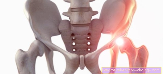
- Child's hip joint (hip dysplasia)
- Baker's cyst
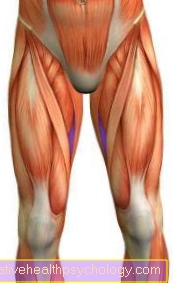
- Soft tissue swelling / hematoma (torn muscle fiber)
- Bursitis
- Achilles tendon tear
- ganglion
- physical therapy
evaluation

Even if the interpretation of ultrasound images seems difficult for the layperson, many diseases can be treated by means of the Ultrasonic be detected. Sonography is very suitable for the detection of free fluids (e.g. Baker's cyst), but also tissue structures such as muscles and tendons can be assessed well (Rotator cuff, Achilles tendon).
The great advantage of this examination method is the possibility of dynamic examination. In contrast to all other imaging procedures (X-ray, MRI, Computed Tomography) can be examined while moving and diseases that only occur when moving can be made visible.
presentation

There are different display methods for the measurement results of an ultrasound examination. They are called Fashion denotes what from the English word for method or proceedings. The first form of application was the so-called A-mode, which is now almost obsolete and only in the Ear, nose and throat medicine for certain questions (for example whether there is secretion in the Sinuses is used. The "A" in A-Mode stands for Amplitude modulation. The reflected echo is received by the probe and plotted in a diagram in which the X axis the penetration depth and the Y axis represents the strength of the echo. This means that the tissue at the specified depth is more echogenic the further up the measurement curve is.
The most common nowadays is B-mode (the "B" stands for Brightness (translated brightness) Modulation) is used. With this display method, the intensity of the echo is displayed using different brightness levels. The individual gray value of an image point therefore reflects the amplitude of the echo at this specific point. A distinction is made again between in the B-mode M-mode and 2D real-time mode. In the 2D real-time mode, a two-dimensional image is created on the ultrasound monitor, which is composed of individual lines (each line is created by a beam sent and received again). Everything that appears black in this picture is (more or less) liquid, displayed in white air, bone and lime.
In order to better assess some tissues, it is useful in some cases to use special Contrast media to use (this method is mainly used for ultrasound in the abdomen).
To that Sonogram to describe, one uses certain terms:
- Anechogenic is called anechoic
- hypoechoic means hypoechoic,
- isoechogenic means echo equal and
- hyperechogenic is called hyperechoic.
The shape of the image visible on the screen depends on the probe used. Depending on which probe is used and how deep the penetration depth is, this process can be used to create up to more than a hundred two-dimensional images per second. The M-Mode (sometimes also called TM Mode: (time) motion) uses a high Pulse repetition frequency (between 1000 and 5000 Hz). In this form of representation, the X-axis is a time axis; the Y-axis shows the amplitude of the received signals. In this way, motion sequences of organs can be represented one-dimensional. In order to obtain even more meaningful information, this method is often coupled with the 2D real-time mode. The M-Mode is particularly common in the context of a Echocardiography used because it allows you to examine individual heart valves and certain areas of the heart muscles separately. Cardiac arrhythmias in the fetuses can also be detected using this method.
Since the beginning of the 21st century there has also been the multi-dimensional echographs: The 3D ultrasound creates a three-dimensional still image. The recorded data are entered into a 3D matrix by a computer and create an image which the examiner can then view from different angles. At the 4D ultrasound (also Live 3D ultrasound called) it is a three-dimensional representation in real time, which means that the three spatial dimensions are added to the temporal. With the help of this method it is possible for the doctor to make movements (for example of an unborn child or of the heart) practically visible in the form of a video.
Doppler sonography
Read more on the topic: Doppler sonography
If you want to get more information (for example about flow velocities, directions or strengths), there are still special procedures based on the Doppler effect: Doppler and color Doppler sonography. The Doppler effect arises from the fact that the transmitter and receiver of a given wave move relative to each other. So if you record the echo that is reflected by a red blood cell, you can use a certain formula to calculate how fast this particle is moving in contrast to the stationary transducer that sent the signal. Color-coded Doppler sonography is even more meaningful, in which normally the color red stands for a movement towards the transducer, the color blue for a movement away from the transducer and the color green for turbulence.
Different organs
Depending on their nature, there are some tissues that can be displayed particularly well with the help of ultrasound, others that can hardly be displayed at all. Tissues that either contain air (such as the lungs, windpipe or gastrointestinal tract) or are covered by hard tissue (such as bones or the brain) are generally difficult to depict.
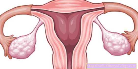
On the other hand, ultrasound provides good results for soft or liquid structures such as the heart, liver and gall bladder, kidneys, spleen, urinary bladder, testicles, thyroid and uterus (possibly including the unborn child). Ultrasound is often used on the heart (heart ultrasound, echocardiography) to examine vessels for any constrictions or occlusions, to monitor pregnancy, to examine the female breast (as a supplement to palpation and mammography), to detect tumors, cysts or Determine organ enlargement or reduction in size of the thyroid gland or to be able to depict organs, vessels and lymph nodes of the abdomen and to detect any tumors, stones (for example gallstones) or cysts that may be present there.
Please also read our pages Ultrasound of the breast and Ultrasound of the testicle, such as Ultrasound of the abdomen
Other areas of application
However, ultrasound is not only used in medicine, it is also used in many other areas of everyday life: for example, not so long ago ultrasound was used to transmit information, for example with remote controls. In addition, you can practically "scan" certain materials with the help of ultrasound, which is used, for example, with sonar to scan the sea floor or with ultrasonic testing devices that can reveal cracks or inclusions in some materials.