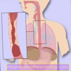
Illustration Baker cyst

- Baker's cyst
(Popliteal cyst) - Femur -
Femur - Semi-membranous muscle -
Semimembranosus muscle - Joint capsule, fiber layer
(yellow) -
Capsula articularis,
Membrana fibrosa - Joint capsule, soft layer
(orange) -
Capsula articularis,
Synovial membrane - Joint cavity
(filled with synovial fluid) -
Articular cavity, synovia - Shin - Tibia
- Internal calf muscle -
M. gastrocnemius, caput mediale - Fibula - Fibula
- Articular cartilage (dark blue) -
Cartilago articularis - Kneecap - patella
- Inner meniscus -
Meniscus medialis
A - Knee joint effusion with Baker's cyst
B - Healthy knee joint
a - swelling in the hollow of the knee
b - swelling in the calf muscles
You can find an overview of all Dr-Gumpert images at: medical illustrations
Related images

Illustration
Knee joint

Illustration
Back of the knee pain

Illustration
Outside knee pain

Illustration
Inside knee pain

Illustration
Pain in the legs

Illustration
Pain in calf