Achalasia
Synonyms
Esophageal spasm, cardias spasm, cardias spasm, narrowing of the esophagus
English:achalasia
Definition of achalasia
The Achalasia is a rare disease that causes a neuromuscular dysfunction (a disorder of the interaction of muscles and nerves) of the esophagus (Esophagus) underlying. The focus is on the lack of relaxation of the lower esophageal sphincter (lower esophageal sphincter) so that the food ingested is not transported properly into the stomach while swallowing. The lower esophageal sphincter usually ensures that the ground food components are in the stomach be transported.
This requires the muscles of the sphincter to relax. In turn, by tensing the muscles, it acts as a valve mechanism to prevent acidic gastric fluid from entering the esophagus (heartburn / Reflux disease).
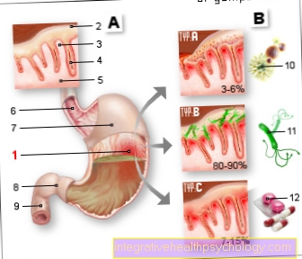
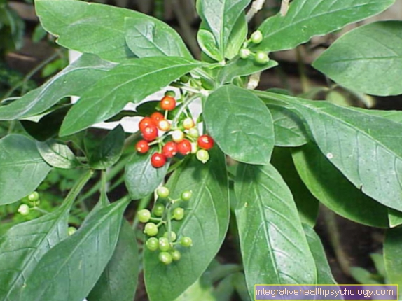
Figure digestive tract

- Throat / throat
- Esophagus / esophagus
- Stomach entrance at the level of the diaphragm (diaphragm)
- Stomach (gaster)
Another characteristic of achalasia is a general poor muscle movement (Peristalsis) of the esophagus during the act of swallowing. The reason for this disease lies in the destruction of the esophageal nerve plexus (loss of nerve tissue of unclear cause = loss / disruption of the Myenteric plexus Auerbach), which regulates the function of the esophagus muscles and is therefore responsible for the harmonious interaction of important muscle groups during swallowing.
Epidemiology
Achalasia is a rare disease (1: 100,000 / year) and usually begins between the ages of 25 and 60. 5% of the patients are children. Men and women are equally affected.
causes
The Achalasia can be divided into two forms:
Primary achalasia:
It is the most common form of the disease. The cause of the development of achalasia is unknown (ideopathic). Viral and autoimmune causes of the disease are suspected.
Secondary achalasia:
Secondary means that achalasia develops as a result of another overriding (primary) disease. In rare cases, a tumor of the esophagus can destroy the nerve plexus of the esophagus (myenteric plexus) and thereby cause achalasia. Even rarer, especially in South Africa, can Chagas disease, be responsible for achalasia. The parasitic pathogen Trypanosoma cruzi attacks the lower part of the esophagus. Here, too, the decline (degeneration) of the nerve cells of the myenteric plexus is characteristic.
Symptoms of achalasia
The signs of illness (Symptoms) achalasia develop insidiously and progress steadily with increasing destruction of the nerve plexus. The dominant symptom is difficulty swallowing (Dysphagia). The dysphagia shows itself in both solid and liquid food. In some cases it is even more pronounced when drinking (so-called. paradoxical dysphagia).
Belching of food occurs (Regurgitation), up to Vomitbecause the food that is swallowed accumulates in the esophagus and is not transported further into the stomach. Characteristically, patients do not complain of a sour taste in the mouth, as in reflux disease (heartburn), because the food does not yet match Stomach acid came into contact.
It can also cause pain, bloating and a feeling of pressure behind Sternum (retrosternal pain) come. This pain can be misinterpreted as heartbreak.
As the disease progresses, patients complain of progression Weight loss, Malnutrition symptoms can occur especially in children.
Achalasia patients often use helpful maneuvers to aid in the act of swallowing and transporting food, such as stretching the neck and back while swallowing.
Complications
A very dangerous complication of achalasia is inhalation of food particles (Aspiration). Patients are particularly at risk at night when the reflexes and thus the gag reflex are weakened. If the inhaled food (Aspirate) in the lower airways, it can become life-threatening lung infection (Aspiration pneumonia) come.
The delayed passage of the food can lead to inflammation processes in the esophageal mucous membrane. In the worst case, such chronic mucosal damage can result Esophageal cancer (Esophageal carcinoma). Achalasia patients have a 15-fold increased risk of developing esophageal cancer.
In the rarest of cases, overcrowding of the esophagus can lead to a tear (Perforation) come in the esophageal wall (Rupture of the esophagus) and for the transfer of food components into the Chest cavity come. Such an event represents an absolute, life-threatening emergency. Apart from bleeding and the injury to other organs during the rupture itself, a life-threatening inflammation of the middle layer can also occur (Mediastinum) of Chest (Mediastinitis) develop.
diagnosis

To secure the diagnosis of achalasia, technical examination procedures are necessary:
X-ray contrast agent examination ("porridge swallow")
This examination is the method of choice for advanced stages of achalasia. In the case of a typical radiographic achalasia finding in the upper esophageal section, a strong enrichment of the esophagus with contrast agent can be seen, as an indication of an excessively dilated esophagus (megaesophagus), followed by a sudden narrowing of the esophagus just before the gastric entrance, caused by the lack of relaxation of the lower esophageal sphincter. The descriptive name for this typical radiological achalasia phenomenon is the "champagne or wine glass" shape of the esophagus.
Esophago gastroscopy (esophagus and gastroscopy)
If the esophagus is narrowed on the "swallow" x-ray, an endoscopy should be performed to rule out other reasons for the narrowing. For example, an esophageal tumor level with the lower esophageal sphincter (esophageal sphincter in front of the stomach) may mimic achalasia. In general, endoscopy (mirroring) is part of routine diagnostics if achalasia is suspected (see Sectsee also: Endoscopy).
Manometry (pressure measurement in the esophagus)
This procedure is particularly suitable for diagnosing achalasia in the early stages. Here, a probe is first placed through the nose of the patient into the stomach and then slowly pulled back towards the mouth. When withdrawing, the pressure in the esophagus is continuously measured by means of a balloon at the end of the probe. A device draws a graphic that shows the pressure conditions in the course of the esophagus. In this way, severe dysfunction of the lower esophageal sphincter (esophageal sphincter) can be diagnosed. Typically, achalasia shows the lack of relaxation of the lower esophageal sphincter during the act of swallowing, as well as an increased resting pressure of the esophagus in this area. Above the esophageal sphincter, the lack of muscle work of the esophagus is evident.