Breast biopsy
What is a breast biopsy?
A biopsy is a diagnostic method in which a material sample is taken from a specific tissue. A breast biopsy is breast tissue. Depending on the suspected underlying disease, different regions of the breast can be biopsied. This usually follows due to a suspected lump in the chest, which should now be examined more closely.
Read more on the subject at: biopsy

Indications
A breast biopsy is usually done when a mass has been detected in a breast. This can be felt in the form of a knot by the affected woman herself or usually by the gynecologist. Even in early detection examinations such as ultrasound or mammography, abnormal regions in the breast can be detected, which should be examined for their entity (benign vs. malignant) by means of a biopsy.
Read more on the subject at: Breast lump
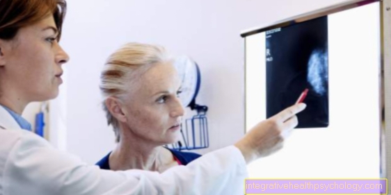
Real indications for a breast biopsy are mainly masses with an unclear origin or a suspicion of malignancy. This is classified according to the BI-RADS criteria, which are used in mammography. A biopsy should be performed if the BI-RADS value is 4 (= suspicious findings, clarification recommended, suspicion of malignancy between 2 and 95%) and a BI-RADS value of 5 (highly suspicious of malignancy over 95%). The highest BI-RADS value is achieved with 6 and denotes breast cancer confirmed by means of a tissue sample. With BI-RADS values below 4, no to low-grade (below 2%) suspicious malignancy abnormalities can be found in the mammography. A biopsy is not recommended in these cases; if necessary, a new imaging of the breast should be done early (e.g. after 6 months) to determine how to proceed.
Read more on the subject at: Mammography
What is a vacuum biopsy?
The vacuum biopsy is a type of tissue removal in which a thin hollow needle is used for the biopsy. The needle is usually inserted into the breast under ultrasound control or controlled by MRI images, from where a tissue sample is drawn directly into the hollow needle. The tissue cylinder obtained from this can then be examined under the microscope.
What is an open biopsy?
With an open biopsy, the tissue is not removed through a fine puncture channel. Instead, the skin over the suspicious area is first opened, then the suspicious tissue is surgically exposed, after which the tissue sample can be taken. Open biopsies are rarely used due to the rather larger intervention; minimally invasive procedures are preferred (if possible). Since the open biopsy is a larger examination with more affected tissue, this is always carried out with local anesthesia, and in major operations even under general anesthesia.
What is a stereotactic biopsy?
In medicine, stereotactic procedures refer to examination or therapy processes in which one acts from several directions. In a stereotactic biopsy, for example, several instruments are aimed at the suspicious area in the chest from different directions. Most of the planning is done on the computer beforehand after evaluating three-dimensional MRI images. Due to the stereotactic nature of the intervention, a precise biopsy of the breast can be performed. This allows you to work very precisely, while at the same time only a small amount of surrounding breast tissue can be damaged by the procedure.
preparation
The preparation of the breast biopsy consists first of all in a detailed diagnosis based on anamnesis, physical examination and imaging (ultrasound, MRI of the breast).The exact method of sampling can then be selected, primarily based on the imaging. Depending on the type of suspected tissue changes, open or closed biopsy samples can be performed. In addition, computer simulations and models can be used to determine which approach is best for the biopsy. Affected people must also be informed in good time before the procedure and of course agree to the examination. Further preparations mainly concern the type of (mostly local) anesthesia carried out.
procedure
The process of the breast biopsy is usually somewhat different, depending on which procedure is chosen. Samples are usually taken under image control. This can be done either using ultrasound or MRI. The place where the biopsy needle is supposed to penetrate the skin must first be disinfected. If multiple biopsies are planned, local anesthesia of the skin and the underlying layers is often performed. In the case of a single biopsy, the sting with the anesthetic syringe would be just as uncomfortable as the sting through the puncture needle, so that in consultation with the person examined, anesthesia is usually not used.
Then, depending on the type of procedure, a vacuum biopsy, a punch biopsy or an aspiration biopsy is performed. All procedures are based on a needle that is pushed into the suspect tissue. A sample then enters the hollow space of the needle through various mechanisms. The tissue sample is then examined microscopically as soon as possible; if necessary, the sample must first be preserved in a tube and sent to a pathological institute. The puncture site itself can usually be closed again with a simple plaster. Only open biopsies are a slightly different procedure, in which the skin and underlying layers have to be surgically removed and, accordingly, have to be properly sutured again after the biopsy has been taken.
How painful is that?
Most breast biopsies are performed using biopsy needles. If only a single sample is taken, it is a one-time needle stick. Since this is no less uncomfortable than an anesthetic injection, local anesthesia is usually dispensed with.
If several biopsies are taken, local anesthesia of the skin and the underlying layers can be performed beforehand. You can feel the needle stick of the anesthetic needle, and the anesthetic can cause some pressure in the tissue. However, the biopsy itself is no longer perceived afterwards. These needle biopsies can cause pain when the anesthesia wears off. However, the pain usually disappears after a few hours to days.
Do you need an anesthetic for this?
Whether or not an anesthetic is performed for the breast biopsy depends heavily on the procedure used. Open biopsies are surgical procedures that are often performed under general anesthesia. Biopsies that are done through needles are either performed under local anesthesia or, in consultation with the person concerned, without anesthesia (if only one or two stitches are required).
Is this possible on an outpatient basis?
Most breast biopsies can be done on an outpatient basis, as either a local anesthetic is used or no anesthetic is used at all. It is also a minor procedure that can usually be performed without complications, so there is no need for medical monitoring after the biopsy. Only open biopsies are usually performed in an inpatient situation, since the affected person should then be observed for longer.
Results
The results of the breast biopsy should initially provide information about whether the changes in the breast tissue are benign or malignant. This can usually be determined using microscopic examinations. This is followed by a precise evaluation of which cells are causing the changes. For example, different cells of the milk ducts or the glands can be affected. A more precise processing of the results usually only takes place if the changes are in need of treatment and / or malignant. In this case, the exact biological responses of the affected cells should be investigated. For example, it can be determined which drugs are particularly effective against the disease, whether hormone therapy is more effective than chemotherapy or radiation.
Read more on the subject at: Benign breast tumor and Tissue samples in breast cancer
Duration until the results
How long it takes to get the results depends on the extent of the biopsy and the local structures. It usually takes a few days before the results of samples that had to be sent to a laboratory are available again. Clinics that have their own pathology department can expect the first results after just a few hours, depending on the urgency. However, the exact biological properties of the affected cells can only be determined through more complex tests, so that these results usually have to be waited for a few days or even weeks.
What does micro lime mean?
Microcalcifications can be detected in the breast using imaging (ultrasound, mammography, X-ray) and a biopsy. Basically, different types of calcifications can appear in the chest. Microcalcifications are areas that measure less than half a centimeter in maximum diameter. While macrocalcifications (larger foci of calcium) are usually benign findings, microcalcifications can speak for both benign and malignant changes, which is why a biopsy is often performed if microcalcifications are found on mammography or ultrasound.
Read more on the subject at: Calcification of the chest
Risks - how dangerous can that be?
The risks of a breast biopsy can be divided into several categories. This can lead to general complications such as bleeding, secondary bleeding, swelling, injuries and inflammation of the skin and subcutaneous fatty tissue. As a result, this can lead to more pronounced and longer-lasting pain in the affected area. An allergic reaction to the local anesthetic and, if necessary, to general anesthesia are also possible.
Complications immediately caused by the biopsy are particularly noticeable in the breast tissue itself. There, individual structures can be damaged or become inflamed, causing long-lasting discomfort within the affected breast. The most dangerous are chest infections that occur after the biopsy. The biopsy creates a puncture channel into deeper tissue regions of the breast. Pathogens (especially skin germs) can penetrate deeper tissue through this channel. As a result, inflammation usually occurs there, which can also spread in the body and thus lead to fever and malaise up to blood poisoning. In general, however, a breast biopsy is a very uninvasive and safe procedure, so serious complications are very rare.
Read more on the subject at: Inflammation of the chest
Duration
Most breast biopsies, including a preliminary examination, disinfection, if necessary anesthesia and needle biopsy, are performed within a few minutes up to half an hour. If a biopsy has to be planned on the computer using three-dimensional images, the preparation usually takes a few days. In this case, too, the biopsy itself is usually performed quickly. Only the open biopsy usually requires a longer period of time, since the beginning and the end of the anesthesia usually take a certain amount of time. Interventions lasting around an hour (depending on how complicated and large the breast surgery are) can also take several hours.
costs
Due to the small procedure, the cost of a breast biopsy is mostly determined by how long the doctor performing the examination takes. In addition, laboratory costs for the examination of the sample material must be calculated. If the biopsy is controlled by ultrasound, the costs remain low; if an MRT device has to be used, it can become more expensive.
Who pays the costs?
The cost of a breast biopsy is usually covered by the health insurance company, so it is unfortunately not possible to provide precise information about the respective prices. Since biopsies are usually only performed for a medical indication, the reimbursement of costs should be fully covered by both private and statutory health insurance.
What are the alternatives?
First, the alternatives to a breast biopsy are imaging tests. Ultrasound, MRI and mammography can be used to assess space requirements. Above all, the probability of a malignant degeneration of the affected region plays an important role. On the basis of various diagnostic criteria, this probability can often be defined very well. If the probability is below 2%, a wait-and-see behavior with follow-up imaging after several months is usually sought. Even if there is a higher risk of dangerous lesions, it is possible to wait and monitor the situation using ultrasound, etc. However, this approach is not recommended in the guidelines.
Read more on the subject at: MRI of the chest and Mammography


