Small intestine
Synonyms in a broader sense
Interstitium tenue, jejunum, ileum, duodenum
English: intestinal
definition
The small intestine is the section of the digestive tract that follows the stomach. This is divided into three sections. It begins with the duodenum, followed by the jejunum and ileum. The main task of the small intestine is to break down the food pulp (chyme) into its smallest components and to absorb these components through the intestinal mucosa.
Illustration small intestine

- Small intestine -
Intestine tenue - Duodenum, upper part -
Duodenum, pars superior - Duodenal
Jejunum junction -
Duodenojejunal flexure - Jejunum (1.5 m) -
Jejunum - Ileum (2.0 m) -
Ileum - End part of the ileum -
Ileum, pars terminalis - Colon -
Intestinum crassum - Rectum - Rectum
- Stomach - Guest
- Liver - Hepar
- Gallbladder -
Vesica biliaris - Spleen - Sink
- Esophagus -
Esophagus
You can find an overview of all Dr-Gumpert images at: medical illustrations
Duodenum / duodenum
anatomy
This section directly follows the exit of the stomach (pylorus). It is approx. 24 cm long, has the shape of a "C" and with this "C" it encloses the head of the pancreas. The duodenum is also divided into an upper part (pars superior), which connects directly to the gastric outlet, the descending part (pars descendens), the horizontal part (pars horizontalis) and the ascending part (pars ascendens).
The duodenum is the only part of the small intestine that is firmly attached to the posterior abdominal wall. In its descending part, the excretory ducts of the bile duct (ductus choledochus) and the pancreatic duct (ductus pancreaticus) end. These usually open together into the papilla vateri (papilla duodenalis major). If the ducts open into the duodenum separately in rare cases, there is an additional pancreatic outlet into a smaller papilla (papilla duodenalis minor).
Further information on the anatomy of the abdominal cavity can be found here: Abdominal cavity

"Internal organs" illustration
- Thyroid cartilage / larynx
- Windpipe (trachea)
- Heart (cor)
- Stomach (gaster)
- Large intestine (colon)
- Rectum
- Small intestine (ilium, jejunum)
- Liver (hepar)
- Lungs or lungs
Venom / ileum
The two longer parts of the small intestine Jejunum) and Ileum (Ileum) lie in the middle of the abdomen and are from the Colon framed. These two sections of the small intestine are very mobile because they rest on a special suspension structure, the so-called Mesentery are hung, which the Intestines flexibly attached to the posterior abdominal wall. This fatty structure also contains the blood vessels, nerves and Lymph nodesthat supply the small intestine. The small intestine is so suspended from the mesentery that it lies in large folds, also called that Small intestinal mesentery are designated.
The jejunum is approx. 3.5 m long, the ileum is approx. 2.5 m. Between these two sections of the small intestine, no sharp border can be drawn with the naked eye. The parts of the small intestine can only be distinguished from each other by means of the tissue (histologically). At the end of the small intestine, the ileum opens laterally into the cecum part of the large intestine, this opening from the large intestinal valve (ileozaekal valve, Bauhinschen flap) is covered. This flap acts as a functional seal between the ileum and the large intestine. The bacteria colonized in the large intestine cannot penetrate into the sterile small intestine through this valve.
length
The small intestine is a very active organ and therefore has no fixed length. Depending on the state of contraction, the small intestine is 3.5 to 6 meters long, with the individual sections being of different sizes. The smallest part of the small intestine is the duodenum (Duodenum), which connects directly to the stomach. It measures an average of 24-30 cm. The is attached to the duodenum Jejunum (Jejunum), which measures 2.5 meters when relaxed. The last section before going into the colon is the Ileum (Ileum), this is about 3.5 meters long. This is Guideline valueswhich can vary from person to person, and from a purely anatomical point of view there is no clear line between empty and ileum.
Wall of the small intestine
Layer structure and structures of the wall of the small intestine
- From the inside, the wall of the small intestine is lined with mucous membrane (tunica mucosa), which is divided into three sub-layers.The top layer is a covering tissue (lamina epithelialis mucosae). In this covering tissue, special cells (goblet cells) are embedded, which are filled with mucus, which they periodically release into the intestinal interior and thus ensure that the intestine can slide. The next lower layer is a connective tissue shifting layer (lamina propria mucosae), followed by a very narrow layer of intrinsic muscles (lamina muscularis mucosae), which can change the relief of the mucous membrane.
- This is followed by a loose shifting layer (tela submucosa), which consists of connective tissue and in which a dense network of blood and lymph vessels runs, as well as a nerve fiber network called the submucosal plexus (Meissner's plexus). This nerve network represents the so-called enteric nervous system and innervates the intestine independently of the central nervous system. In this layer of the duodenum there are also the so-called Brunner's glands (Glandulae interstinales), which form various enzymes and an alkaline mucus, which the Stomach acid able to neutralize. The following intestinal muscle layer (tunica muscularis) is divided into two sub-layers, the fibers of which run in different directions: first an inner, strongly developed circular muscle layer (stratum circulare) and then an outer longitudinal muscle layer (stratum longitudinal). Between this ring and the longitudinal muscle layer runs a network of nerve fibers, the myenteric plexus (Auerbach plexus), which innervates (stimulates) these muscle layers. These muscles ensure the wave-like movement of the intestine (peristaltic movement).
- Another connective tissue layer (Tela subserosa) follows.
- The conclusion is formed by a covering of the peritoneum, which lines all organs. This coating is also called tunica serosa.
Mucous membrane from the small intestine
The small intestine needs a large area to absorb the food components. By a severe wrinkling and numerous protuberances, a great increase in the surface of the mucous membrane is achieved. This is guaranteed by various structures:
- Kerkig folds (Plicae circulares)
These are ring folds that form the rough relief of the small intestine and in which both the mucosa and the submucosa protrude. - Small intestine villi (Villi interstinales)
These finger-shaped protrusions, 0.5-1.5 mm in size, are found in all sections of the small intestine, in which the epithelium and the lamina propria protrude. - Lieberkühn crypts (Glandulae interstinales)
In the valleys of the villi there are tubular depressions that extend to the lamina muscularis. - Microvilli
This so-called “brush border” forms the micro-relief of the small intestinal mucosa and enlarges it ten times. In the case of the microvilli, the cytoplasm (filling content of the cells) of the individual small intestinal cells (enterocytes) is everted.
The fine tissue (histological) differences between the individual small intestine sections are briefly presented here:
- Duodenum
The duodenum is characterized by very high kerking folds and leaf-shaped imposing small intestinal villi. The most important feature, however, are the Brunner's glands (Glandulae interstinales), which occur only in the duodenum, are located in the submucosa and participate in the formation of the small intestinal juice and form enzymes such as maltase and amylase. - Jejunum
Here, the kerking folds become smaller over time, the small intestinal villi become longer and have a more finger-shaped structure - Ileum
The kerking folds are particularly low in this section of the small intestine and are completely absent in the lower ileum. The villi of the small intestine also become shorter and shorter and the number of goblet cells increases as the intestine progresses. The large number of lymph follicles (accumulation of lymph cells) in the ileum is particularly noticeable. If many follicles are gathered in one place, this place is also called Peyer's plates. These structures are involved to a large extent in the immune defense of the intestine.
Function / tasks
As part of the digestive tract, the main role of the small intestine is the Further processing of the edible pulp and the Absorption of the contained nutrients, electrolytes, vitamins and fluids.
In the small intestine, the previously chopped up food components are broken down into their basic components and absorbed. This is done on the one hand by the Addition of digestive enzymes to chyme, on the other hand through contact of the basic components with the cells of the small intestinal mucosa. The small intestine uses several tricks to design the contact surface of the chyme with the mucous membrane and thus the absorption of the food as large as possible: Wrinkled protuberances protrude into the interior of the intestinal sections, from which cell assemblies such as tentacles protrude again. Every single cell of these tentacles now has so-called again on its surface Microvilli, finger-like protuberances that enlarge the contact area again. Overall, the small intestine enlarges its surface so on up to 200 m².
If the chyme reaches the duodenum through the stomach passage, the secretions from the gallbladder and pancreas are emptied in its so-called "descending part". The pancreas produces up to 1.5l secretion daily. This consists largely of bicarbonate, which the neutralizes the acidic milieu of the porridge.
The main work is done here, however, by the ones that are also included pancreasenzymes, they further break down the food. There is a specific enzyme for every component of food: for Fats (including pancreatic lipase and phospholipase A), carbohydrates (alpha amylase), proteins (including trypsin and aminopeptidases), DNAComponents (Ribonuclease, deoxyribonuclease) etc.
The important part of the bile for digestion is the Bile acidswhich have a special property. They can bind both fat and water and thus simplify the processing of fats in food. The bile acids, which are synthesized from cholesterol, form so-called fats with the food Micelles. These are small "lumps" of fat, consisting of the fat components inside and the bile acids as a protective ring to the watery external environment.
The mixture of chyme and digestive enzymes is now through the Small bowel peristalsis transported further towards the colon. The walls of the small intestine contract the more slowly the further they move away from the stomach. The Duodenum contracted 12 times per minutewhile that Ileum only 8 contractions per minute having.
The sections of the small intestine differ not only in the number of contractions per minute, but especially in their Wall construction and the absorbed food components. In the duodenum, mainly calcium, iron, Magnesium, Single and double sugar absorbed.
The further course will now be in descending order fat soluble Vitamins, Egg whites, water-soluble vitamins and Fats reabsorbed until the bile acids in particular are reabsorbed in the terminal ileum and vitamin B12 is absorbed.
The further towards the colon you move, the more accumulations of Lymph follicles can also be found in the intestinal wall. The intestine not only serves as a digestive organ here, but also as a Immune defense station against germs and bacteria ingested with food.
The final part of the small intestine forms the Bauhin'sche flap. It defines the transition from the small to the large intestine and prevents the stool from flowing back from the large to the small intestine. From the Bauhin'schen flap the number of Intestinal bacteria rapidly and the species that occur change.
Movement / peristalsis
After inclusion in the Small intestinal mucosa the nutrients are carried into the bloodstream. Through the vascular network (capillaries) in the small intestine villi, the sugars, the amino acids (from peptides) and the short to medium-chain fatty acids are absorbed into the blood vessels and passed on to the liver via the portal vein. The long-chain fatty acids, the cholesterol esters and phosphilipids, are broken down into large protein-fat molecules (Chylomicrons) and via the lymphatic vessel in the small intestinal villi, past the liver and into the blood flow funneled.
The intestines are also important for them Absorption of water. Approx. 9 liters of fluid are absorbed in a day. Of this, about 1.5 liters come from the liquid drunk and the rest are the liquids (secretions) that the Gastrointestinal tract forms. This includes saliva, Gastric juice, small intestinal juice, pancreatic and biliary juice.
Ingestion

After being absorbed into the mucous membrane of the small intestine, the nutrients are transferred into the bloodstream. Through the vascular network (capillaries) in the small intestine villi, the sugars, the amino acids (from peptides) and the short to medium-chain fatty acids are absorbed into the blood vessels and passed on to the liver via the portal vein. The long-chain fatty acids, the cholesterol esters and phosphilipids, are broken down into large protein-fat molecules (Chylomicrons) built in and channeled through the lymphatic vessel in the small intestinal villi, past the liver and into the bloodstream.
The intestine is also important for the absorption of water. Approx. 9 liters of fluid are absorbed in a day. About 1.5 liters of this comes from the liquid drunk and the remainder are the liquids (secretions) that the gastrointestinal tract forms. These include saliva, gastric juice, small intestinal juice, pancreatic and biliary juice.
Figure digestive tract

Digestive tract
A. - Food route
a - digestive organs
in the head and neck
(upper part of the digestive tract)
b - digestive organs
in the body cavity
(lower part of the digestive tract)
- Oral cavity - Cavitas oris
- Tongue - Lingua
- Sublingual salivary gland -
Sublingual gland - Trachea - Trachea
- Parotid gland -
Parotid gland - Throat - Pharynx
- Mandibular salivary gland -
Submandibular gland - Esophagus - Esophagus
- Liver - Hepar
- Gallbladder - Vesica biliaris
- Pancreas - Pancreas
- Colon, ascending part -
Ascending colon - Appendix - Caecum
- Appendix -
Appendix vermiformis - Stomach - Guest
- Large intestine, transverse part -
Transverse colon - Small intestine - Intestine tenue
- Colon, descending part -
Descending colon - Rectum - Rectum
- Nach - anus
You can find an overview of all Dr-Gumpert images at: medical illustrations
Small intestine pain
Pain in the small intestine is not easy to pinpoint. There are many different conditions that can cause pain in the small intestine. The spectrum here ranges from simple blockages or Gastrointestinal inflammation up to heavier ones chronic inflammation up to Intestinal ulcers or Mesenteric infarcts.
Many of these diseases also cause relatively unspecific pain in the lower abdomen, which on the one hand cannot be easily distinguished from one another and also resemble pain patterns in other diseased organs such as the pancreas, gall bladder, peritoneum or large intestine.
Pain in the small intestine shows up depending on the clinical picture different "pain qualities". These range from colic-like (severe, rippling) pain when the small intestine is blocked (Ileus) from dull, long-lasting pain to acute, stabbing pain in an ulcer or an ulcer acute inflammation.
In principle, the motto here is that the more acute and stronger the pain, the more serious the disease is. It should also be noted whether, in addition to the pain, a so-called Defense tension occurs here, which is reflective and can only be triggered to a limited extent arbitrarily Hardening of the abdominal wall means when touched.
Pain in the small intestine area must always be seen in the context of known previous illnesses. For example, pain in acute small intestinal inflammation after gastrointestinal viruses or food poisoning can be "normal" as long as it does not last longer than four days, on the other hand, for example, expresses itself Mesenteric artery infarction with following Reduced blood supply the affected section of the small intestine with short, severe pain, which then improves and almost disappears, while the disease takes on threatening proportions.
Inflamed small intestine

The inflammatory disease of the small intestine is called Enteritis designated. Due to the close positional relationship, the stomach and large intestine can also be inflamed, and these forms of disease then become gastroenteritis (Stomach) or Enterocolitis (Colon) called.
Enteritis is classified according to various criteria: 1. Is the enteritis infectious or non-infectious 2. Is the inflammation acute or chronic? 3. What caused the inflammation?
Infectious enteritis can be caused by bacteria (including salmonella, shigella, E. coli, clostridia), viruses (including rotaviruses, noroviruses, adenoviruses) or parasites (including amoebas, worms, fungi).
Non-infectious enteritis refers to inflammation of the small intestine that is of medicinal origin (cyclosporine, cytostatics), is triggered by radiation therapy, is a result of insufficient blood supply in the corresponding section, is caused by toxins, is caused by allergies such as food allergies or after operations or is idiopathic ( without a known cause) are like ulcerative colitis or Crohn's disease.
Enteritis mainly manifests itself as diarrhea, which is often accompanied by nausea and vomiting. Other, more unspecific symptoms are intestinal cramps, abdominal pain and fever. In the course of the disease, the increased excretion of water and decreased absorption lead to signs of dehydration and disturbances of the electrolyte balance such as dizziness, tiredness, listlessness and leg cramps.
The therapy of enteritis depends on its triggers. Most enteritis heals spontaneously, with diarrhea subsiding within 3-7 days and nausea and vomiting subside within 1-3 days. In these cases, treatment is symptom-oriented and depending on the degree of severity, possibly with medical treatment of nausea, diarrhea and electrolyte imbalance. In the case of more persistent inflammation, a detailed discussion with the patient is important in order to clarify the above-mentioned triggers, and pathogens are also detected via stool samples. Therapy is then adapted to the results of the examinations. Bacterial and parasitic enteritis, for example, are treated with antibiotics if the symptoms persist.
Major illnesses
Ulcerative colitis
Ulcerative colitis is also a disease belonging to the group of inflammatory bowel disease (IBD). Ulcerative colitis is particularly characterized by involvement of the large intestine, but can sometimes also affect the small intestine. One then speaks of an "ingrown" inflammation of the small intestine ("Backwash ileitis"). This disease is also triggered autoimmunologically and causes abdominal pain and bloody ones diarrhea (Diarrhea) noticeable.
Further information on this topic can be found at: Ulcerative colitis
These inflammatory bowel disease (IBD) can theoretically affect the entire gastrointestinal tract from the oral cavity to the anus. However, the disease preferentially affects the lower small intestine (terminal ileum) and often appears with symptoms such as cramp-like abdominal pain and slimy diarrhea (diarrhea). The characteristic of this autoimmune disease, however, is the segmental infestation of the intestinal mucosa.
Further information on this topic is available at: Crohn's disease
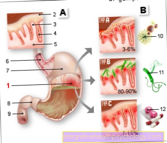
Duodenal ulcer
The so-called duodenal ulcer refers to an ulcer in the duodenum. The two main causes of this very common disease are bacteria Helicobacter pylori and pain medication like aspirin or Not-S.teroidalA.nti-R.heumatic (NSAIDs). A dangerous complication of ulcer disease occurs when the ulcer reaches a larger vessel causing life-threatening bleeding (Gastrointestinal bleeding) is coming.
Celiac disease
This condition is commonly known as gluten-sensitive enteropathy, or native sprue. This is an intolerance of the mucous membrane of the small intestine to the adhesive protein (gluten) found in many types of grain. Those affected complain of diarrhea and weight loss. The therapy for this disease is lifelong gluten free diet.
Further information on this topic can be found at: Celiacia


