The human bloodstream
definition
The bloodstream is made up of the heart and blood vessels. The heart serves as a pump to pump the blood that is in the vessels through the body. For this purpose, the human body has a vascular system which, starting from the large vessels that arise directly from the heart, branches out further and further in order to reach every part of the body.
When the blood reaches the “end”, for example at the fingertips, toes or the organs, it flows back to the heart to close the circulation, to be “recycled” again and to be redistributed in the body.
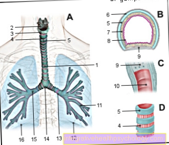
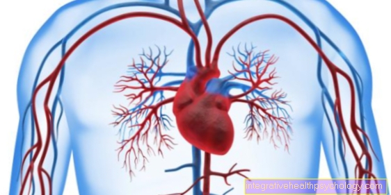
Illustration of the blood circulation

Human blood circulation
A - pulmonary circulation
(small cycle)
Right HK> Lung>
Left HK
B - body circulation
(big cycle)
Left HK> Aorta> Body
red - oxygenated blood
blue - deoxygenated blood
- Neck-head-vein -
Brachiocephalic vein - Superior vena cava -
Superior vena cava - Right atrial -
Atrium dextrum - Right ventricle -
Ventriculus dexter - Right lung -
Pulmodexter - Lower vena cava -
Inferior vena cava - Common pelvic vein -
Vena illiaca commonis - Clavicle artery -
Subclavian artery - Aortic arch - Arcus aortae
- Left atrium -
Atrium sinistrum - Left ventricle -
Ventriculus sinister - Left lung -
Pulmo sinister - Abdominal aorta -
Abdominal aorta - Femoral artery -
Femoral artery
HK = ventricle
You can find an overview of all Dr-Gumpert images at: medical illustrations
Functions of the bloodstream
The task of the bloodstream is to supply the organs with all the nutrients they need to carry out their respective functions. The blood takes over this transport of nutrients.
The blood transports oxygen through the body to all organs, because these cannot work without oxygen and would die. In addition, the carbon dioxide generated in the organs is absorbed and transported away by the blood. The oxygen "swims“Not just floating around in the blood, but is bound to a transport medium called hemoglobin during transport.
One molecule of hemoglobin (imaginable as a large sphere) four molecules of oxygen (imaginable as small balls) bind to itself and release it again elsewhere, absorbing carbon dioxide. You could compare it to a drinks delivery company with a car (hemoglobin) four boxes of water (Oxygen for survival) to a household (organ) brings home and four empty boxes of water (Carbon dioxide that has been consumed) takes back with him to make room for the new, full ones. The beverage supplier takes them to his company (lung) to replenish them there.
Other nutrients, such as fats, sugar or proteins from food, are also transported by the blood and absorbed from the blood by their target organ.
Waste products that arise in the organs, such as urea, are absorbed by the blood and transported to their excretory organ.
In addition, messenger substances (hormones) are distributed in the bloodstream, which ensure that signals (for example hunger) can be passed on within the body.
Another task of the blood circulation is to regulate the temperature in the body. The blood can absorb and release heat so that a constant state can be established. The cells that are responsible for our blood to clot when we are injured are also transported in the bloodstream.
Vascular system
The beginning of the vascular system can be imagined like a tree. Starting with the thick one aorta (Diameter: 2.5 - 3.5 cm) the vessels branch out further and further and become thinner the further they are from Heart are away.
The vessels can be divided into Arteriesthat carry the oxygen-rich blood from the heart to the whole body. In this way the blood will increase nutrient and oxygen withdrawn so that the oxygen-rich blood becomes oxygen-poor blood. This deoxygenated blood gets over the Veins directed back to the heart.
The transition between arteries and veins is formed by the Capillaries. These are the smallest vessels with a diameter of 5-10 µm through which just one can enter red blood cell (Erythrocyte) fits through. Because these vessels are so narrow, the blood flows through them very slowly. So here is a lot of time for the organs to absorb oxygen from the blood and at the same time produce it Carbon dioxide to give to the blood.
The capillaries are then followed by the Veins. Here the size curve is exactly the opposite of that of the arteries. Starting with small veins that connect to the capillaries, these become thicker and thicker until the largest veins flow into the heart.
Classification of the bloodstream
The blood circulation is divided into a large circulation, the body circulation, and a small circulation, the pulmonary circulation.
To understand these two cycles, one must first look at the structure of the heart. The heart consists of two heart chambers (Ventricle) and two atria (Atrium).
The left atrium and left ventricle are also grouped together as the left heart, and the right atrium and right ventricle as the right heart. The atria and ventricles on one side are each separated by valves, the so-called leaflet valves. These valves only open when the heart beats. Otherwise they are closed so that the blood does not flow back.
In the great circulation, starting from the left ventricle, in which there is oxygen-rich blood, this blood is released during a heart action (Heartbeat) is pumped into the subsequent aorta. For this, the blood has to pass through the aortic valve, which is opened by the pressure and otherwise closed. From here the blood can reach the entire body and all organs. As described above, the blood flows from there via the veins back to the heart.
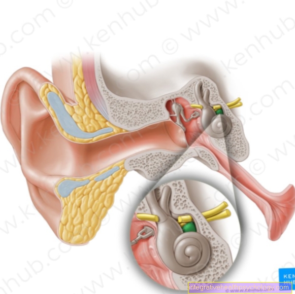
The connection to the heart is made by the largest veins, the upper and lower vena cava (Venae cavae superior and inferior), which open into the right atrium from above and below.
The upper vena cava has previously collected the venous, i.e. the oxygen-poor blood from the head and neck region, the lower vena cava that from the body. So here, on the right side of the heart, is deoxygenated blood. The blood is drawn from the right atrium through the tricuspid valve (right AV valve) is pumped into the right ventricle. Since the blood is low in oxygen and nutrients, it must first be re-enriched with oxygen and nutrients before it can supply the body with them again. This happens in the small circulation, the pulmonary circulation.
The pulmonary circulation starts from the right ventricle. From there the venous blood is drawn into the pulmonary artery (Pulmonary artery), which is closed by the pulmonary valve when at rest. The pulmonary artery carries the blood to the lungs so that nutrient enrichment can take place there. There is also a vascular system in the lungs that is made up of arteries, capillaries and veins, just like in the body's circulation.
The arteries in the lungs, which continue to branch out, are accompanied by the bronchi, which carry the air out of the airways. The exchange of substances takes place in the smallest vessels, in the capillaries, as this is where the lowest flow velocity is achieved. The capillaries are separated by a minimally thin wall from the end sections of the airways, the alveoli (Alveoli), Cut. Over that thin wall (membrane) substances can migrate in both directions. Here, oxygen is absorbed from the alveoli into the blood and, on the other hand, carbon dioxide is released from the blood into the alveoli so that it can be exhaled. The blood, which is now rich in oxygen again, is then released via the pulmonary veins (Pulmonary veins) directed back to the heart.
Here four pulmonary veins (two on each side) open into the left atrium. From there they are pumped through the mitral valve (right AV valve) into the right ventricle of the heart, from where they are fed back into the great circulation, the body's circulation. In contrast to the right heart, the left heart contains the oxygen-rich blood.
Circulatory diseases
Blood circulation disorders are particularly common in older people.
One of the most famous diseases is arteriosclerosis. This is a change in the innermost vascular layer in the small arteries. Cholesterol and calcium deposits increasingly narrow the blood vessel and prevent adequate blood flow in the structures it supplies.
This leads to circulatory disorders, for example peripheral arterial occlusive disease (PAOD), which often manifests itself in a reduced blood supply to the legs. Affected patients then have increasing discomfort when walking.
If atherosclerosis affects the arteries supplying the heart (coronary arteries), in extreme cases this can lead to a heart attack, as this is then not supplied with sufficient oxygen. If the arteries that lead to the brain are narrowed, this can lead to a stroke.
In children and adolescents, most circulatory disorders can be traced back to heart defects.