Adipose tissue necrosis
definition
Adipose tissue necrosis is understood to be the destruction of adipose tissue through cell death (necrosis) of adipose tissue cells (adipocytes), which can affect various organs and parts of the body. Necrosis means that cells within a living organism die. In adipose tissue necrosis, fat cells die and release the stored fat, which is absorbed by the surrounding connective tissue cells. Encapsulation results in cysts filled with an oily liquid, so-called oil cysts. Calcification processes within the capsule form hard knots in the tissue, which can be up to several centimeters in diameter.

causes
There are many causes for the development of adipose tissue necrosis.
The most common cause is trauma, i.e. tissue damage caused by external violence (contusion, bruise). Blunt force (e.g. belt injuries caused by an impact in a car accident) directly damages the fat cells. As a result, the fatty tissue dies necrotic and oil cysts form, which can be felt as hard nodules. Blood vessels can also be severed, whereby the tissue is undersupplied and also damaged. The latter can also occur during operations and minor surgical interventions.
Another cause can be improper injections of cytotoxic drugs that damage adipose tissue and cause it to die.
If you have an acute inflammation of the pancreas (Pancreatitis) the protein lipase reaches the surrounding tissue, is activated and then destroys fat cells. Lipase is an enzyme that normally enters the small intestine with the pancreas and breaks down dietary fats there. The pancreas can also be damaged by external force or circulatory disorders, which also causes lipase to escape and destroy fatty tissue.
In rare cases, for reasons that have not yet been clarified, fatty tissue necrosis can develop, especially in the area of the lower legs. The necrosis becomes visible as reddish papules on the skin, which turn brown over time and remain painless. This clinical picture is called necrobiosis lipoidica diabeticorum, since diabetics are more often affected.
Read more on the subject at: Symptoms of diabetes
After an injection
In the event of an incorrectly performed injection or infusion, the liquid does not get into the punctured vessel but into the surrounding tissue (extravasation). This leads to painful swellings and fluid build-up in the affected area. Such unintentional extravasations are usually harmless and the fluid in the tissue is quickly absorbed and removed by the body.
However, the wrong injection of certain drugs, especially cytostatics, can lead to tissue necrosis. Cytostatics are toxic substances that are used in chemotherapy to treat cancer and kill tumor cells. If the drug gets into the adipose tissue, the fat cells are killed and adipose tissue necrosis occurs.
Even diabetics who need insulin often develop fatty tissue necrosis in the abdominal wall as a result of the frequent subcutaneous injections of insulin.
With cortisone
Cortisone has an anti-inflammatory effect and cortisone-containing syringes are therefore administered, among other things, for pollen allergies, hay fever and orthopedic problems in the buttocks area. If the cortisone is not injected deep enough into the muscle or if fluid flows back from the puncture channel into the fatty tissue, the fatty tissue dissolves and dies. The necrotic fat tissue is visible on the skin as deep dents that can be several cm2 in size. In some cases the tissue can renew itself and the dent will disappear after a few months.
Read more about this: Cortisone Injection - Indications for Use and Side Effects
After irradiation
Radiation therapy is an effective method of treating cancer patients. The local radiation destroys tumor cells and thus increases the chances of recovery. However, healthy adipose tissue in the vicinity of the tumor can also be destroyed, causing adipose tissue necrosis and oil cysts to form in the irradiated tissue. This is a benign finding because the necrosis does not have any risk of degeneration and therefore does not need to be treated.
After an operation
Adipose tissue necrosis can develop after an operation. The cell death of fat cells in the course of necrosis can then lead to the formation of oily cysts or fat-filled cavities, which over time become more and more calcified. These calcified cysts can then usually be felt as a swelling or lump under the skin.
After a breast reduction
A breast reduction is based on a surgical procedure in which tissue is removed. The incisions made during the operation can also lead to the destruction of fat cells or fat tissue necrosis.
The adipose tissue necrosis may show up as swelling in the breast. Occasionally the tissue around the dead cell material is inflamed, which leads to tenderness when touching the affected chest area. The skin over the necrotic chest area can also be red and thickened. Furthermore, there may be swelling of lymph nodes in the immediate vicinity.
Read more on the subject at: Breast reduction
diagnosis
Doctors diagnose adipose tissue necrosis by feeling the lumps under the skin. Adipose tissue necrosis is actually harmless and does not need to be treated further, but cannot be distinguished from malignant growths by means of a tactile finding alone. Therefore, the further diagnosis is made through an ultrasound examination, although there can often be difficulties in differentiating it from a carcinoma, especially in the breast. In order to be able to definitively rule out a possible tumor, the lump must be surgically removed and the tissue must be finely healed by a pathologist (histologically) can be examined under the microscope.
You might also be interested in that: Importance of the biopsy for breast cancer diagnosis
What do you see in the ultrasound?
In addition to a palpation examination, which usually shows the fatty tissue necrosis as a lump, imaging methods such as ultrasound can also be used. Benign changes or swellings have typical characteristics in the ultrasound, including fatty tissue necrosis.
These include a clear demarcation from the environment - the dead or damaged tissue takes up space, but adjacent cells are not destroyed. Furthermore, the benign changes show a smoothly delimited and homogeneous or uniform structure. The tissue can also be easily compressed (pressed in) by the transducer.
Concomitant symptoms
Necrosis of the fatty tissue is usually unproblematic and does not cause any discomfort or pain. The necrosis leads to the formation of hard nodules of different sizes, depending on how much fatty tissue has gone under. In rare cases, the tissue around the necrosis can become inflamed and cause pain.
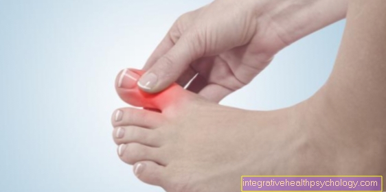
Lymph nodes that drain lymph from the affected area (e.g. in the armpit region in the case of necrosis in the breast) can swell and be easily palpable. There may be a bluish discoloration of the skin over the lump. In some places (especially on the thighs and buttocks), fatty tissue necrosis causes deep and very large dents in the skin, which are perceived as very annoying by the patient and can even lead to reduced self-esteem or inferiority complexes. In such cases, surgery can provide relief for aesthetic reasons.
Pain in adipose tissue necrosis
Adipose tissue necrosis is usually not associated with pain. Occasionally, an inflammatory reaction around the dead area of fat cells leads to a sensation of pain. This can be particularly noticeable when touching the skin over the affected cell area.
Treatment / therapy
As a result of the necrosis, nodes form from dead fatty tissue, which are always benign, do not cause any symptoms and therefore usually do not require treatment. If the lumps become inflamed and cause pain, they can be surgically removed.
The problem with diagnosing by means of the tactile findings is that it is not possible to differentiate between a lump caused by the loss of adipose tissue and a malignant tumor. Cancer can only be clearly ruled out through a fine needle biopsy, in which cells are removed from the node with a thin cannula and then examined under a microscope, or the complete removal of the node.
Duration
Adipose tissue necrosis generally shows a very good course without pain. If the necrosis is not located in the chest, it usually does not have to be removed and patients can live with it without any symptoms.
In the breast, a clarification must be carried out to clearly rule out a carcinoma. The necroses, which are encapsulated from the rest of the fatty tissue, usually last a lifetime and only in very rare cases go away on their own.
For aesthetic reasons, deep dents in the skin can be surgically removed or injected with fat, which are particularly common in fatty tissue necrosis on the buttocks and thighs.
forecast
The adipose tissue necrosis usually has a good prognosis. In most cases no therapy is necessary. In a few cases, however, removal may be advisable, for example, with fatty tissue necrosis in the breast.
Surgical removal may be necessary here if no clear distinction can be made between adipose tissue necrosis and a possible malignant tumor using imaging techniques. To be on the safe side, the lump or tumor is then removed and the removed tissue is examined, which then enables a differentiation.
Traumatic adipose tissue necrosis
Traumatic adipose tissue necrosis occurs, for example, when adipose tissue is crushed or bruised. This can happen during an accident, for example. As with surgery, this can damage adipose tissue with subsequent destruction of fat cells and the development of adipose tissue necrosis.
In the case of adipose tissue necrosis, regardless of its origin (surgical interventions, radiation, traumatic adipose tissue necrosis), no therapeutic measures are usually necessary. The necroses are often palpable as lumps under the skin, but regardless of this, they can usually be regarded as harmless.
cyst
When fat cells die as a result of trauma or surgery, the fat can liquefy and lead to the formation of so-called oil cysts. These are cavities that are filled with liquefied fat and often calcified over time. These are mostly benign changes that usually do not require therapy.
Predestined body regions
On the chest
Adipose tissue necrosis is very common in the breast, as the breast is mainly made up of fat cells in addition to glandular and connective tissue. An operation on the breast (e.g. breast-conserving therapy (BET) for breast cancer, breast reduction or the insertion of silicone implants) can destroy fat cells or the blood supply can be disrupted by severing vessels. This leads to the necrotic death of breast fatty tissue and the formation of oil cysts, which can be felt as solid knots from the outside. The formation of fat necrosis in the breast can also be caused by the insertion of faulty silicone implants. The implants can tear open and the contents of the prosthesis leak into the surrounding tissue and destroy the cells.
Another common cause of adipose tissue necrosis on the chest is blunt injuries from trauma when the tissue is crushed or bruised. As a rule, fatty tissue necrosis in the chest is not a problem, but the injury can also tear blood vessels, free droplets of fat get into the bloodstream and vessels, e.g. clogging in the lungs (fat embolism). Such a severe course and complications are very rare.
On the thigh
Adipose tissue necrosis forms on the thigh as a result of injuries, bruises or incorrectly performed injections.
In the area of the thigh and the abdominal wall, so-called Marcumar necrosis can occur as a rare side effect of long-term therapy with Marcumar. Marcumar is an anticoagulant drug that is used to thin the blood. The increased tendency to clot at the beginning of the treatment can clog small capillaries and lead to vascular occlusion. The consequences are circulatory disorders and the destruction of fatty tissue with the formation of oil cysts.
On the buttocks
In the area of the buttocks, incorrectly placed injections often lead to the formation of fatty tissue necrosis, which can be recognized by deep dents in the skin. Necrosis is mainly caused by repeated administration of cortisone depot syringes. The drug must be injected deep into the muscle, as an injection that is too superficial leads to the destruction of the fatty tissue and large dents can appear in the affected area between the buttocks and hips.


.jpg)