MRI examination
Synonyms
- Magnetic resonance tomography
- Magnetic resonance imaging
- Magnetic resonance examination
English
- NMR (nuclear magnetic resonance)
- MRI (magnetic resonance imaging)
Radiation exposure during an MRI examination
The advantage of a Magnetic resonance imaging in contrast to Computed Tomography and roentgen is that there is no radiation exposure for the patient.
The MRI scans arise when a strong magnetic field is generated, which affects the hydrogen atoms in the human body. These then send out radio waves to different degrees, depending on what type of tissue it is. These waves are made by one computer detected and processed into sectional images.
Since there is no radiation exposure, there are no known side effects from an MRI examination. For young patients in particular, an MRI is a good method to visualize the soft tissues without the risk of radiation exposure.
Do you have to be sober for an MRI scan?
Usually the patient does not have to appear sober for a magnetic resonance imaging scan. Eating and drinking is allowed in advance.
An exception is the examination of certain organs in the abdomen (MRI abdomen). Before an MRI scan of the intestines, bile, or stomach (please refer: MRI of the stomach) For example, the patient should be sober so that the images can be easily assessed. During these examinations, it is also often necessary to drink a contrast medium before the examination.
Whether or not the patient has to come to the examination on an empty stomach will be communicated beforehand.
Duration of an MRI examination
The Duration of a magnetic resonance imaging examination is variable. Depending on which area is to be mapped and how many images are to be recorded, it takes shorter or longer.
However, the examination itself usually takes about 15 to 30 minutes. Then there is the preparation time and the waiting time.
Part of the preparation is to remove all metal parts that are on the body or clothing. In addition, the patient must be placed on the examination table and special pillows may have to be used to fix the examined body part.
If the administration of contrast medium is necessary, the examination takes longer, as this is usually injected into the arm vein after the first pass before a second pass starts.
Basics and technology

The Magnetic resonance imaging, also as Magnetic resonance imaging (MRI) is a modern cross-sectional process that makes use of the principles of so-called nuclear magnetic resonance. Unlike, for example, the Computed Tomography are used to generate the images no x-rays (please refer roentgen), but rather strong magnetic fields and radio waves.
With the help of this MRI examination slice images of almost any part of the body in any angle and direction can be generated in a non-invasive manner (without interfering with the body) in a relatively short time.
This information is available in digital form, which enables the radiologist to generate various views of the examined body part after the examination with the help of powerful computers.
Technical and physical basics
The central core of a MRI - Investment (Magnetic resonance imaging) is a superconducting electromagnet weighing tons, mostly cooled with liquid helium. Transmitting and receiving antennas are built into its inner wall.
If necessary, additional antenna coils are connected to the magnetic resonance tomography depending on the body region to be examined. For special examinations there are specially shaped coils, e.g. B. for the examination of the head, the knee joint, the Spine or the (female) chest (MR mammography). So that the examination is not disturbed by other radio waves, the MR examination room is through a Faraday cage shielded.
The human body is made up of innumerable tiny biological magnets due to the abundance of hydrogen protons. This is used in magnetic resonance tomography. Due to their rotation (Nuclear spin) These hydrogen protons develop a magnetic moment and the protons behave like small magnetic gyroscopes that align themselves in an externally applied strong magnetic field based on the field lines of the magnetic field.
The Magnetic resonance imaging (MRI examination) essentially takes place in three steps:
First a strong, stable, homogeneous magnetic field of 1 - 3 Tesla is generated around the body (10,000 - 30,000 times stronger than the earth's magnetic field) and thus a stable alignment of the protons is achieved.

As a second step the MRI examination this stable alignment is changed by using electromagnetic high frequency energy in the form of a radio signal at a specific angle to align the Hydrogen protons irradiates. The hydrogen protons are set in motion by the radio signal from the MRI. After the radio pulse has been switched off again, the hydrogen protons return to their original position and in the process give off the energy that they absorbed from the radiated radio pulse. In the third step, the energy emitted is generated by receiving coils (Principle of antennas) measurable. Thanks to a sophisticated arrangement of these receiving coils, one can three-dimensional coordinate system Measure exactly where and when which energy has been released. The measured information is then converted into image information by powerful computers.
Above is an example of one open MRI (Magnetic resonance imaging).
MRI of the different parts of the body
MRI of the head
In the MRI image of the head you get an image of all the structures in the head. It allows statements about that skull, the structure of the brain, the arterial and venous blood conductors of the head, as well as other cavities and soft tissue of the head. By recording a magnetic field in the MRI as opposed to the CT or roentgen Examination, the patient experiences no radiation exposure.
As with most MRI examinations, the patient drives into the tubular tomograph on a couch. In a head examination, the head and the upper body are accordingly in the device. The patient must lie still during the exposure, otherwise the images may be blurred.
The recording takes between 20-30 minutes. During this time, the device can sometimes make loud, knocking noises, which the patient shouldn't be put off by. The examination is not harmful for harmful and is used to diagnose many diseases. Above all, the structures of the brain, for example for tumors, infarcts or inflammatory changes, are examined.
MRI of the knee
Magnetic resonance imaging is also the best means of imaging soft tissue in the knee.
The tubular device, which has a round opening on two sides, is also used. The difference to some other MRI examinations is that the patient does not have to go inside the tube with his upper body. A common problem is a Claustrophobia at the MRI In an MRI examination of the knee, the patient only has to go into the tomograph up to about the hip. The patient should be prepared, for example by going to the toilet, as the examination takes about 30 minutes.
For the knee, too, the MRI examination is the more expensive, but the less harmful alternative to the CT. The ligament and cartilage structures of the knee can be made visible in a particularly detailed manner. Hence it is useful in diagnosing Damage to the menisci, of the Cruciate ligaments, of the Collateral ligaments and the cartilage.
An MRI is also recommended for unexplained knee pain that has not found a cause using any other diagnostic method.
MRI of the heart
As a supplement to the usual heart diagnostics, for example the Ultrasound of the heart, magnetic resonance imaging of the heart can be performed as a non-invasive procedure. It depicts the soft tissue structures in the chest with great detail. For example, the size of the heart chambers, the structure of the heart muscle and, above all, the coronary arteries can be represented very precisely.
The MRI examination of the heart has gained in importance, especially in the last decade. Because an MRI recording takes a long time, heart examinations are often fuzzy due to the constant movement of the muscle. Thanks to new devices and methods, the images can be recorded more quickly and the heart movements can be compensated for in the image. It cannot examine patients with pacemakers or other implants in the body. The patient has to go through the tomograph with his entire upper body.
The MRT examination is particularly suitable to detect infarcts or blockages of the coronary arteries. It is also used at Suspected heart muscle inflammation and for examination after heart surgery.
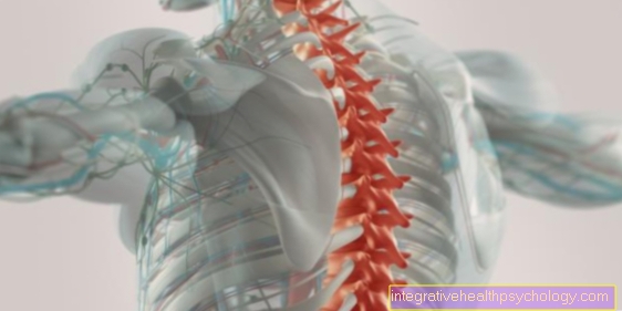
MRI of the cervical spine
The MRI examination of the cervical spine is used more and more frequently for a wide range of diseases. It depicts all structures of the spine, including intervertebral discs and nerves, in great detail. The high contrast of the image makes it possible to diagnose even minor changes in the spine or nerve compression. The MRI also shows inflammation and tumors well, which is why it is particularly used for diagnostics.
During the MRI scan of the neck, the patient must first go into the tomograph with his head. Here, too, he lies quietly on a lounger that automatically moves into the interior of the tube. Like most MRI examinations, the recording takes between 20 and 30 minutes. The patient can be given a contrast medium for a more precise diagnosis. As with all examinations that work with a magnetic field, such as the MRI, he is not allowed to wear any metallic objects on or in the body. All types of prostheses or pacemakers are included.
MRI of the prostate
The MRI examination of the prostate is often a necessity in diagnostics, since the prostate is often difficult to examine with conventional methods due to its location in the pelvis.
Sometimes it also becomes Preventive examination of the prostate performed as the Prostate cancer is the most common male cancer. The risk of developing prostate cancer increases steadily with age.
An exact diagnosis cannot be made by palpation and blood tests alone.
Due to the lack of radiation, the MRI examination is a health-friendly alternative to computed tomography. An MRI image is also much more accurate, but it takes about 20-30 minutes and usually costs more. For a more detailed examination, a contrast agent is injected into the patient beforehand via a venous access.
The MRI examination of the prostate is used primarily for prevention in patients at risk, suspected carcinomas and for interventions such as biopsies or operations. If the patient has metallic implants or prostheses in their body, they are unfortunately out of the question for an MRI examination.









.jpg)
