Skull base
definition
The skull base is used in anatomical terminology Base cranii called and is part of the neurocranium. The skull (lat. Cranium) is in the Viscerocranium (Facial skull) and that Neurocranium (Brain skull) divided.
Classification

The base of the skull is inserted into the Basis cranii interna, the side facing the brain and the Base cranii externa, the outside divided. In the rear part of the outer skull base, this also reshapes the contours of the brain; in the front part, however, the outer skull base is involved in the formation of the nasal cavities and the hard palate.
Basis cranii interna
The inside of the skull base can be turned into three skull pits which are divided from one another in stages. One distinguishes the Anterior cranial fossa, media and posterior, i.e. the anterior, middle and posterior fossa.
Anterior cranial fossa
The anterior fossa is mainly from Frontal bone (Frontal bone) and the Sphenoid bone (Sphenoid bone) formed. The Ethmoid bone (Ethmoid) has only a small part of the anterior cranial fossa. The anterior cranial fossa supports and forms the frontal lobe of the cerebrum Roof of the eye socket, in addition, she is involved in the formation of the Nasal cavity involved. The olfactory threads from the olfactory bulb, which is located in the anterior cranial fossa, pull through the ethmoid bone Nasal mucosa. The anterior cranial fossa has at the transition to the middle cranial fossa with the canalis opticus a passage point for the optic nerve and for the arteria ophthalmica, which the eyeball supplied with blood.
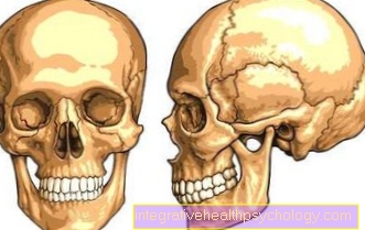
Figure skull bones

- Frontal bone -Frontal bone
- Parietal bone - Parietal bone
- Occiput - Occipital bone
- Temporal bone - Temporal bone
- Sphenoid bone - Sphenoid bone
- Ethmoid - Ethmoid bone
- Zygomatic bone - Os zygomaticum
- Nasal bone - Nasal bone
- Upper jaw - Maxilla
- Lower jaw - Mandible
- Tearbone -Lacrimal bone
- Chin hole - Mental foramen
- Ploughshare - Vomer
- Under eye cavity hole -
Infraorbital foramen - Zygomatic arch -
Arcus zygomaticus - Temporomandibular joint -
Articulatio temporomandibularis - External ear canal -
Meatus acousticus externus - Mastoid process
(Part of the temporal bone) -
Mastoid process - Lambda seam -
Sutura lambdoidea - Cuticle seam -
Sutura squamosa - Crown seam - Coronal suture
- Orbital upper edge -
Margo supraorbitalis
You can find an overview of all Dr-Gumpert images at: medical illustrations
Fossa cranii media
The middle fossa consists of a right and a left part, both of which are constructed in exactly the same way, and houses the two temporal lobes of the cerebrum. Most of the bony structure consists of the Temporal bone (Temporal bone) and additionally from the Sphenoid bone (Sphenoid bone). The pituitary gland, the hormone-producing pituitary gland, is located in the sella turcica (Turkish saddle) of the sphenoid bone. The cranial nerves III, IV, V1 and VI pull through a gap in the sphenoid bone, the fissura orbitalis superior, to the eye muscles in order to innervate them by motor. In addition, a nerve runs from the middle cranial fossa to supply the upper jaw (Maxillary nerve) through the foramen rotundum and a nerve supplying the lower jaw (Mandibular nerve) through the foramen ovale. In the middle cranial fossa there is also the carotid canal as a passage point for the internal carotid artery, which supplies the brain with blood.
Posterior cranial fossa
In the formation of the posterior fossa is mainly that Occipital bone (Occiput) involved that Temporal bone (Temporal bone) and that Sphenoid bone (Sphenoid) have smaller parts of the bony structure. The upper part of the posterior fossa houses the Occiptal lobe of the cerebrum, in its lower part the cerebellum. In the bones of the posterior fossa there are imprints of the Sine, of the venous blood conductors of the brain. In addition, the largest passage point for the base of the skull, the foramen magnum, is also found here. The Foramen magnum houses the medulla oblongata and blood vessels, it connects the posterior fossa with the spinal canal. With the porus acusticus internus (inner auditory canal), there is also the passage for the eighth cranial nerve, which is responsible for the sense of hearing and balance, in the posterior fossa.
Base cranii externa
The outer side of the base of the skull with its anterior part forms the Choans, the rear openings of the Nasal cavities. Also the upper jaw and the bony palate belong to the outside of the skull base. In the rear area of the outer skull base are the Occipital condyle (Occipital condyles), which are connected to the first vertebral body.
Injuries
A Skull base fracture is a potentially life-threatening injury that often occurs in traffic accidents. The base of the skull breaks depending on the direction of the applied force along certain fault lines, these fault lines are particularly thin areas of the bony structure. In the area of the anterior cranial fossa is the bone plate of the Ethmoid bone (Ethmoid) very thin, which is why fractures are particularly common in accidents here. A consequence of a bone fracture in this area can be a tear in the dura tough Meninges, as this is fused to the bone in this area. Another consequence can be Loss of smell by tearing off the olfactory threads, as these pull through the ethmoid bone at this point and can be injured if a bone breaks. With this so-called frontobasal fracture it often occurs Exit from Liquor (Cerebral fluid) from the nose, as the cerebrospinal fluid can no longer be held back due to the tear in the hard meninges and now runs out of the nose. Also, blood vessels can be injured and Blood running out of your nose. In the case of a lateral fracture of the base of the skull, the meninges and vessels in the central fossa can be injured, blood and liquor then run out of the ear on the affected side. Other symptoms of a skull base fracture include severe headache, or vomiting Impaired consciousness.