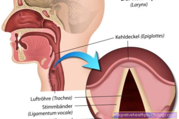
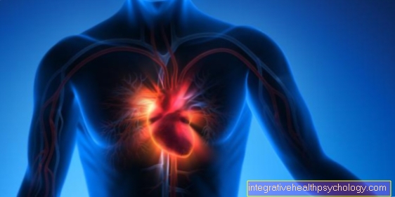
How do you recognize myocarditis?
introduction
An inflammation of the heart muscle (Myocarditis) is a serious illness that can be fatal if it is not recognized in time. As the symptoms are usually very unspecific, early diagnosis is often a challenge. Typical symptoms include tiredness and decreased resilience that occur during or after an infection. Blood samples are also examined to ensure rapid diagnosis. Technical examinations such as an EKG, cardiac ultrasound or MRI also play an important role in detecting myocarditis.

What are the symptoms of myocarditis?
Heart muscle inflammation is very difficult to recognize because the symptoms are non-specific. Even experts usually need technical aids to diagnose the disease. It is important to know that heart muscle inflammation usually occurs as a result of an infection. This can take the form of a common cold, but the flu can also cause myocarditis.
The signs of myocarditis are usually only increased tiredness and a decrease in performance, as the heart cannot pump enough power when it is exerted. It is also characteristic that the symptoms persist after the infection. Some sufferers feel a palpitations in which some heartbeats suddenly become very noticeable. This can be an expression of a heart rhythm disorder caused by myocarditis. Pain in the chest area is rare. These are only typical for heart muscle inflammation if the pericardium is also affected.
Read more on this topic at: Symptoms of myocarditis
How does the doctor diagnose myocarditis?
Doctors usually rely on technical equipment to diagnose myocarditis. Nevertheless, the anamnesis is first in the foreground. On the one hand, the doctor asks for the symptoms. Symptoms such as tiredness, reduced resilience, possibly also cardiac arrhythmias and pain in the chest area would be typical. These complaints generally indicate a heart problem. In order to make the diagnosis of myocarditis, it is also asked whether the person concerned had an infection in the past. Since colds or flu can trigger inflammation, this aspect is particularly important and effective.
The physical exam may reveal water retention. These occur when the heart's pumping capacity decreases. Heart murmurs can also be heard in some people. After the examination, an EKG is first written, in which cardiac arrhythmias can be found. In addition, blood samples are examined in which enzymes from destroyed heart muscle cells can be found. A search for the pathogen (virus, bacterium) is also possible. The next steps in the diagnosis include imaging tests such as an X-ray, a cardiac ultrasound or a cardiac MRI. To confirm the diagnosis, a biopsy is usually taken from the heart muscles.
Read more on this topic at: Diagnosis of myocarditis
Which laboratory values / blood values indicate a heart muscle inflammation?
The most important laboratory values for heart muscle inflammation are the so-called heart markers. These are enzymes that are normally only found in heart muscle cells. If these cells are destroyed, the enzymes enter the blood. So they can only be detected in the laboratory if there is a heart problem. Typically, the CK-MB and troponin are examined here. The causative agent can also be found in the blood. Viruses are usually the cause of myocarditis, but an infection with bacteria is also conceivable. Since the illness is usually based on a cold or flu, the general inflammation levels in the body are also increased, especially CRP.
Read more on this topic at: Inflammation of the heart muscle - blood tests
CRP
CRP is an abbreviation for C-reactive protein. It is therefore a protein that is released in the body in response to inflammation and infection. Heart muscle inflammation is characterized on the one hand by the local inflammation of the heart muscle cells, on the other hand the disease is accompanied, at least at the beginning, by an infection that generally increases the inflammation values. Therefore, it is not possible to diagnose myocarditis based on an increase in CRP alone. On the other hand, a high CRP value can be an indication that the heart muscle is inflamed.
What you might also be interested in on this topic: Levels of inflammation in the blood
CK-MB
CK MB stands for "creatine kinase muscle brain". This is an enzyme that is only found in heart muscle cells. If these muscle cells are destroyed, for example by an inflammation of the heart muscle, the creatine kinase enters the bloodstream and can be measured there. However, an increase in the CK-MB value does not necessarily indicate an inflammation of the heart muscle. Other causes can be a heart attack or increased stress on the heart muscles.
Troponin
Troponin T and I are usually mentioned together with the heart-specific enzymes, but they are actually structural proteins. So it is about protein chains that belong to the structural part of the heart muscles. They have their function in the contractile (contracting) area of the heart muscle cells and are primarily responsible for converting electrical signals from the excitation conduction system into a mechanical contraction of the muscle cells. Troponin T and I occur exclusively in the heart and therefore specifically indicate damage to the heart muscles. If the cells and with them the surrounding structures are damaged by an illness, their components get into the blood and can be determined there.
Read more on this topic at: Troponin
What does the heart ultrasound show?
Heart ultrasound, also known as echocardiography, has the advantage that it can be carried out very quickly and easily in acute situations. In this way, a first impression of the condition of the heart can be obtained within a very short time. Based on the assessment of the ultrasound examiner, further diagnostic procedures or an immediate initiation of therapy can then be ordered.In the case of an inflammation of the heart muscle, the heart echo is usually used to rule out other cardiac causes (originating from the heart). The inflammation itself can only be recognized in a few cases, which is why a heart ultrasound often shows an inconspicuous finding.
However, some unspecific signs may indicate an inflammation of the heart muscle. This includes a restricted function of the left ventricle. This can be recognized, for example, from the fact that the heart muscles in the affected area do not contract sufficiently. In addition, a larger part of the blood that enters the left ventricle is not pumped out of the heart. Regional wall movement disorders can also be detected. They also indicate a malfunction of the heart muscles. If the heart muscle inflammation is accompanied by an effusion in the pericardium, this can be seen as a border around the heart.
Read more on this topic at: Echocardiography
What does the EKG show in the case of myocarditis?
Heart muscle inflammation usually has very unspecific symptoms, some of which are specific to the heart. Since the symptoms are different, the ECG can also be very different. Disturbances of the heart rhythm are particularly noticeable in the ECG. Therefore, forms of myocarditis that are associated with cardiac arrhythmias are particularly easy to recognize. This rhythmic disturbance can take on all forms from a changed beat speed (tachycardia = too fast heartbeat) to complicated arrhythmias. The arrhythmias can affect both the atria and the ventricles or both together. A distinction between the various forms is possible using an EKG.
Other abnormalities in the EKG are similar to those of a heart attack. This can lead to a reduction in the distance between the S-wave and the T-wave. One speaks of a so-called ST depression. It is also possible to negate the T-wave. Other disturbances of the excitation conduction can also be found in the EKG. For example, if the electrical signal is no longer passed into both chambers of the heart, it is called a bundle branch block (if the right heart chamber is no longer activated, there is a right bundle branch block).
Also read: EKG for an inflammation of the heart muscle
Does an MRI of the heart make sense in case of myocarditis?
An MRI of the heart is useful if there is already a suspicion of myocarditis. The severity of the disease can be better assessed with the help of an MRI. Above all, disorders of the pump function and the movements of the heart wall can be examined in detail. The force that the heart can muster is also measured in an MRI. In this way, the entire functionality of the heart muscles as well as the functionality of individual wall sections can be assessed. Thus, MRI is not the first choice in diagnostics. However, the need for different therapeutic measures can be assessed more precisely with the MRI.
Read more on this topic at: MRI of the heart
Can I tell if I have myocarditis by listening to it?
It is almost impossible to diagnose myocarditis simply by listening. This is due to the fact that often no special features are noticed during the examination. If something suspicious is found, it is usually a question of very unspecific signs, which can also have another cause. Heart muscle inflammation is often associated with cardiac arrhythmias. When listening, these can be found mainly in the systole (the tension phase of the heart). Pericardial rubbing (the rubbing of the two leaves of the pericardium) may also be heard if the pericardium is affected by the inflammation.
How can you recognize myocarditis in children?
In many cases, it is more difficult to detect myocarditis in children than in adults. Heart muscle inflammation occurs after about five percent of viral infections. Since children are particularly susceptible to infections and colds, they are at greater risk of developing myocarditis. Therefore, in children, particular attention should be paid to heart-specific symptoms. In children in particular, the disease is quite mild in the early stages, so that it is often indistinguishable from a simple cold. One should pay attention if the children are still limp, tired and less productive even after the infection has subsided. If the disease is not recognized in time, permanent damage to the heart can occur. Therefore, especially after infections with a fever, you should take a break from exercise of about a week. If a child is suspected of having myocarditis, the child goes through the same diagnostic steps as any adult patient. The signs are the same: tiredness and reduced performance, possible occurrence of cardiac arrhythmias, water retention, etc.