coccyx
Synonyms
Coccyx, Os coccygis
introduction
From an evolutionary point of view, the tailbone is a developmental artifact. It is considered to be a remnant of the Tail human ancestors.
From an anatomical point of view, the tailbone of an upright person forms the lower section of the spine that points towards the floor. In addition to the cervical, thoracic, lumbar and sacral vertebrae, the tailbone thus represents the last part of the spine.
Although it consists of firmly fused sub-units, it plays an important role in various movement sequences. This fact is due to the fact that the coccyx serves as the point of attachment or origin of various ligaments and muscles of the pelvic area. In particular, the structures of the pelvic floor and the hip joint are attached to the coccyx through connections.

anatomy
The actual tailbone is made up of approximately four to five individual coccyx vertebrae together. However, these vortices are through what is called a Synostosis to a single fused uniform bones.
In this context, the term synostosis describes a condition in which two bony structures that previously only covered cartilage or connective tissue were connected to one another, merged with one another over time. Compared to the vertebral bodies of the cervical, thoracic or lumbar spine, almost all typical anatomical features have disappeared in the area of the coccyx.
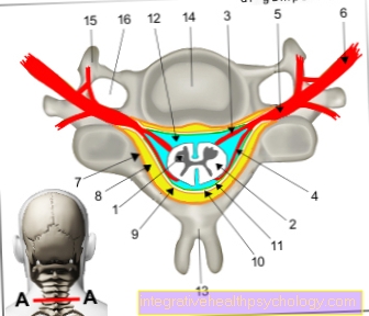
Figure tailbone and lumbar spine

Lumbar spine (blue)
- First cervical vertebra (carrier) -
Atlas - Second cervical vertebra (turner) -
Axis - Seventh cervical vertebra -
Vertebra prominent - First thoracic vertebra -
Vertebra thoracica I - Twelfth thoracic vertebra -
Vertebra thoracica XII - First lumbar vertebra -
Vertebra lumbalis I - Fifth lumbar vertebra -
Vertebra lumbalis V - Lumbar cruciate ligament kink -
Promontory - Sacrum - Sacrum
- Tailbone - Os coccygis
You can find an overview of all Dr-Gumpert images at: medical illustrations
Diseases of the coccyx
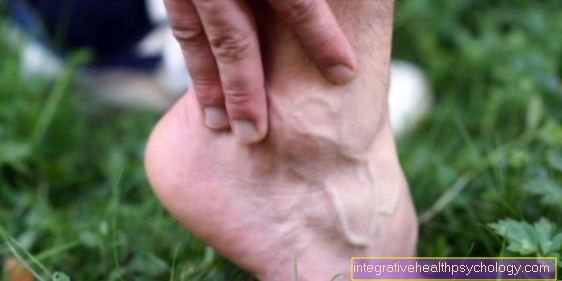
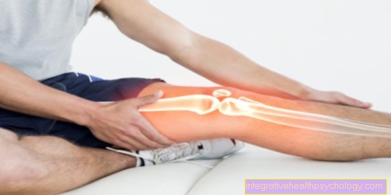
The tailbone is particularly at risk when falling on the buttocks. In addition, trauma in the area of the coccyx is often caused by direct forces (e.g. a kick) against this bony structure.
Both classic fractures and dislocations are among the most common diseases of the coccyx. Characteristic of trauma to the terminal bone of the spine are severe pain, sometimes lasting for weeks, which is aggravated when sitting or walking. Coccyx pain when sitting is the pain that occurs in the last and lowest section of the spine when sitting. Affected people try to minimize the pain stimulus that occurs usually by shifting weight to one side of the pelvis.
Taking pain medication may relieve symptoms for the time being. Paracetamol® (PCM for short) or Ibuprofen® are particularly suitable for the acute treatment of pain in the coccyx area. However, since the problems become noticeable again quite quickly after the pain medication has ended, if there is pain in the coccyx, a doctor should be consulted and an appropriate diagnosis initiated.
Coccyx dislocation

Dislocations of the coccyx are compared to dislocations of the hip or Shoulder joint quite seldom. In most cases, the presence of a dislocation of the coccyx is not even diagnosed as such.
Affected patients suffer from severe pain that makes normal sitting almost impossible. Treating coccyx dislocation is basically pretty straightforward. After a successful diagnosis, the attending physician will put the index finger in the Rectum introduce. Then the bone must be gripped from the rectum with the inserted finger and fixed. The practitioner's thumb should be used externally during the entire procedure print exercise on the tailbone. To release the dislocation and return the tailbone to its original position, it must be slightly removed with the index finger Sacrum be pulled away. At the same time, the sacrum should be pressed towards the feet.
If the reduction is successful, an immediate decrease in pain symptoms can be expected. If there is no corresponding pain relief, it can be assumed that the tailbone is still in a dislocation position and the treatment was therefore unsuccessful.
Furthermore, there is the possibility that there are no problems in the area of the coccyx in spite of the symptoms typical of dislocation. If severe pain is triggered to the left or right of the sacrum during the repositioning attempt, this indicates that the person concerned is ill Sacroiliac joint Near.
Tailbone fracture

Even with fractures of the coccyx (tailbone fracture), the affected patient usually feels severe pain that sets in quickly. Furthermore, a fracture of the coccyx is clinically indicated by the appearance of bruises (technical term: hematoma), which clearly emerge while sitting.
While a simple bruise or a dislocation can be diagnosed and, if necessary, treated in the course of a digital rectal examination, the fracture of the tailbone is often a challenge. The diagnosis of fractures in the region of the tailbone can be made by taking x-rays.
Immobilizing such a fracture is almost impossible due to the localization. In most cases, the fracture of the tailbone is treated with painkillers. Since the affected patients mostly suffer from very severe pain, higher doses of painkillers can be taken.
In addition, a so-called ring pillow can be used to relieve pressure while sitting. The pressure created by the body mass is then not directed to the tailbone but increasingly to the gluteal muscles.
If the pain symptoms do not subside within a few weeks despite these measures, surgical treatment of the fracture may be necessary. The fracture of the tailbone can be fixed during the surgical procedure. In extremely rare cases, however, the tailbone end piece must be completely removed.
Coccyx fistula

A coccyx fistula is a chronic inflammatory disease that occurs in the area of the gluteal folds.
Basically, it does not come from the bony tailbone, but is more often caused by hair that has penetrated the skin. Nevertheless, coccyx fistulas can also be caused by severe bruises or congenital malformations of the coccyx.
Clinically, such a fistula is manifested by the sudden appearance of severe pain, swelling and redness in the area of the gluteal folds. In addition, the affected patients suffer from extreme pressure sensitivity in some cases. In the case of very advanced coccyx fistulas, the emergence of bloody or purulent secretions from the opening of the fistula can be observed regularly. If such a fistula is present, surgical treatment is the treatment of choice. Such an operation can be performed either on an outpatient or inpatient basis.
A distinction is made between different stages of the coccyx fistula:
- The so-called bland form is a mild form of the coccyx fistula and does not show any signs of inflammation. However, the opening of the fistula is rarely recognizable on the skin.
- The acutely abscessing coccyx fistula is suppurated because it has become inflamed (usually due to thick hair, sweat, rubbing of clothes, etc.).
- The third stage is the chronic coccyx fistula, which does not show any acute signs of inflammation, but causes discomfort due to the constant secretion of blood and pus and itching. It can often only be detected by blood / pus stains in the underwear.
As a prophylaxis, especially after an operated coccyx fistula, if it is known that there is a tendency to form a coccyx fistula, the hair in the region should be removed by laser epilation so that the hair is destroyed right down to the roots. The region should always be kept hair-free after the operation by shaving thoroughly and regularly.
You can also find more information under our topic: Coccyx fistula
Surgery for a coccyx fistula
In order to treat the coccyx fistula successfully, a surgical opening of the fistula duct and thus a Operation absolutely necessary. Other forms of treatment are currently not considered to be successful.
In the classic surgical method the coccyx fistula is usually stained with methylene blue. The tissue marked in this way is then removed over a correspondingly large area. The operation will go down to that coccyx cut and the Periosteum scraped off there so that recurrences can be reliably avoided.
The operation is performed under general anesthetic, but in less severe cases it may also be under local anesthesia be performed. In severe cases, that one may be necessary inpatient hospitalization is adhered to for up to four days. Usually, however, the operation is now carried out on an outpatient basis.
The generous excision (cutting out) of the coccyx is the classic therapy of the coccyx.
However, there is also an alternative minimally invasive surgical techniques, such as the coccyx sinus surgery Karydakis or that Pit picking according to Bascom.
These surgical techniques are compared to the classic variant painless surgical techniques. You become partial endoscopically performed and are significantly more complex than the classic surgery of the coccyx fistula.
There are surgical techniques with Flap sculptures (Limberg plastic, rhomboid plastic, V-Y plastic) who work with dislocated skin flaps. They are very complex, but if carried out successfully, they can ensure a more aesthetic appearance of the wound area and the healing rates are significantly more successful.