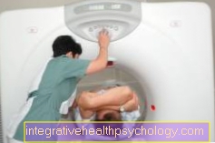
Radiation exposure of computed tomography

In computed tomography, radiation leads to a high level of radiation exposure. Compared to X-rays, this radiation exposure is particularly high and accordingly more dangerous than an X-ray examination.
Nevertheless, computed tomography (short CT) has many advantages over X-rays. On the one hand, cross-sectional images of the body can be made, on the other hand, organs and soft tissues are shown much better than would be possible with X-rays.
Due to the high radiation exposure, one often tries magnetic resonance imaging (short MRI) to evade. Magnetic resonance tomography can also produce sectional images of the body without any radiation exposure. However, depending on the recording, it takes a long time to get an image using magnetic resonance imaging. Computed tomography, on the other hand, only takes a few milliseconds.
In addition, with computed tomography contrast media can be injected into the vein, which then makes the demarcation between two organs or two tissues even better. Nevertheless, there is always the risk of high radiation exposure in the computer tomograph.
Each patient receives an average radiation dose of around 4 mSv per year (mSv = millisievert, the unit in which the radiation dose, i.e. radiation exposure, is given). If a patient now receives a whole-body CT, i.e. a picture of his entire body using computed tomography, this corresponds to a load of 10-20mSv. This means that the radiation exposure from a single computed tomography exposure exceeds the average annual value by 3-5 times. For this reason, a full-body image using computed tomography is only taken in very rare cases, for example when looking for a tumor focus but could not find it using magnetic resonance therapy.
On the other hand, a CT of the abdominal cavity (abdomen) made. Here the radiation exposure is (Radiation exposure) 8.8-16.4 mSv. This corresponds to twice to four times the dose of radiation that a patient normally receives within a year "collect“Would.
The radiation exposure is not quite as high when the Rib cage (thorax). The radiation exposure from the computed tomography recording is here 4.2-6.7mSv. This is roughly equivalent to a patient's annual dose.
Often a computed tomography image of the Lumbar spine carried out, especially in patients with a suspicion of one disc prolapse. The radiation exposure is approx 4.8-8.7mSv. But because of the alternative of MRI, a CT should be carefully considered in the case of a herniated disc.
The information on radiation exposure always fluctuates considerably, as it depends on how strong or how thin a patient is. For a particularly obese (thick) People have to use a higher radiation dose and thus a higher radiation exposure so that the radiation can also get through the fat to the organs.
Already 4 kg Obesity mean a significantly higher radiation exposure. In the case of slim people, on the other hand, the rays can penetrate directly to the organs without major obstacles, so the radiation dose does not have to be particularly high.
head

Especially for investigations of the Head computed tomography is often used. The advantage is that especially with one stroke (Apoplexy) or if there is bleeding into the brain by a Ruptured veins or arteries this is recognized within a few seconds.
The disadvantage is, as always with computed tomography, radiation exposure in and on the head. An examination of the head leads to a comparatively low radiation exposure with only 1.8-2.3mSv. That corresponds roughly to the radiation exposure for half a year.
Desire for children
A recording using computed tomography always leads to a high level of radiation exposure. Therefore, during pregnancy, a Computed Tomography just in an absolute emergency must be carried out, as it is still not known what the effects on the unborn child would be.
The exception is a computed tomography of the head, which has fewer effects on the unborn child.
A patient cherishes one Desire for children and if you have to undergo an examination by means of computed tomography, this is in principle not a problem.
However, it is important that Ovaries and uterus be protected from radiation exposure by computer tomography, as the desire to have children may otherwise remain unfulfilled.
The problem is that the radiation exposure in our gonads (Gonads), i.e. in Testicles in men and in the ovaries (Ovary) is one of the largest.
It is therefore important to have a CT scan of the abdomen (abdomen) Shield the gonads as much as possible so that the radiation exposure during the computed tomography does not destroy the desire to have children.
For the man there are therefore so-called examinations by means of computed tomography Testicular capsules. These capsules are placed around the testicles and shield them so that they are not exposed to any radiation. The nurses or the doctor often point out the possibility of shielding with a testicular capsule, but if they do not, the patient should not hesitate and ask about it.
For women, on the other hand, it is more difficult because the gonads of women, namely the ovaries, are in the body. There is therefore a small one for women Lead apronthat is placed over the ovaries. This lead apron ensures that at least most of the rays are kept away and that no excessive radiation exposure stands in the way of having children.
Sinuses
Computed tomography is also often used to examine the Sinuses used. Since the entire head is usually x-rayed, there is a radiation exposure of approx 1.8-2.3mSv. This corresponds roughly to the radiation exposure for half a year.
cancer
With computed tomography, there are sometimes very high levels of radiation that put an enormous strain on the body. Therefore it is important that a patient be exposed to this radiation must agree and should be informed about the risks beforehand. In addition, the so-called Risk-benefit assessment. The benefit of the investigation should always outweigh the risk.
Whether the radiation exposure to computed tomography cancer It is difficult to say because it is not known whether the cancer, which appeared years after treatment, was caused by radiation exposure or not. It can be due to radiation exposure too Skin changes come, but these then occur immediately after exposure to radiation.
The radiation also causes changes in the DNA of the irradiated cells. This can lead to so-called strand breaks, loss of base and many other changes in the DNA. These then cause the cell to multiply differently than it did before or to perish.
Normally, such errors are corrected by the body's own enzymes. However, it can also be that the flaw in the DNA is due to radiation exposure irreparable is. In this case, the computed tomography and the resulting radiation exposure can lead to cancer. It is therefore always important to weigh the risk against the benefit of the examination.








.jpg)




.jpg)


