Examination of the small intestine according to Sellink
How does the investigation work?
The Sellink examination method is also known as enteroclysis or double-contrast examination of the small intestine according to Sellink. It is used to depict the small intestine and thus to identify various intestinal diseases.
The patient must first be sober and have taken laxative measures, otherwise the intestine cannot be assessed.
In the course of the examination, the patient is given two different contrast media.

A positive contrast agent (Barium sulfate) and a negative contrast agent (Methyl cellulose).
In this context, positive means that the contrast agent leads to an increase in the signal in the imaging, that is, the areas on which the contrast agent attaches appear lighter.
In this case they are Intestinal walls.
The negative contrast agent, on the other hand, leads to a weakening of the signal intensity. A double contrast is obtained.
First, the patient is given a probe inserted through the nose, through which the positive contrast agent is administered first.
This is followed by the negative contrast agent, which ensures that the positive contrast agent is distributed over the intestine and adheres to the intestinal walls. The negative contrast agent with the weaker signal intensity is then in the middle of the intestine, so that the Intestinal walls shine brighter than the lumen. This ensures that the intestinal walls can be assessed particularly well on the patient's X-ray images.
The doctor pays attention to them Folding and thickness of the intestinal walls, on Leaks, i.e. whether the contrast agent emerges at one point Intestinal motor disorders, on Constrictions (Stenoses) of the intestine as well as filling defects, i.e. places where no contrast agent accumulates.
Of the Small intestine can therefore be examined very well for abnormalities during the investigation.
Indications for the Sellink examination
There are various indications for the Sellink examination method for which it is used.
This includes the inflammatory bowel disease (Crohn's disease, Ulcerative colitis), Disorders of the intestinal motor skills, the detection of tumor diseases of the intestine, Diverticula, such as Abscesses, Fistulous ducts and Constrictions (Stenoses).
However, there are also circumstances in which the Sellink examination method may not be used.
This includes the suspicion of a leak in the intestinal walls (perforation), as otherwise the contrast agent would escape into the free abdominal cavity.
In the case of barium sulfate, this would lead to serious complications with inflammation of the peritoneum, which is difficult to treat and dangerous for the patient.
In addition, the Sellink examination method must not be used if the patient has been in the last 14 days operated on the abdomen was suspected of having a Paralysis of the intestines (paralysis) or one Intestinal obstruction (Ileus) consists.
Examination according to Sellink in the MRI
Usually the investigation method after Sellink is under X-ray control carried out.
Alternatively, however, you can also use the MRI be performed.
This procedure is particularly popular with people for whom radiation exposure through x-rays should be avoided. For example at Children or young people.
The procedure is otherwise the same as for the X-ray-supported examination.
The patient is given two different contrast media via a probe. Instead of Barium sulfate is usually used during an MRI scan Gadolinium used.
Contrast media can also be administered via the vein. This then accumulates in the area of possible Foci of inflammation so that the doctor can draw conclusions about the activity of an inflammatory process.
Very active foci of inflammation shine more strongly on the MRI images than less active foci.
However, the Sellink examination method using MRI is not suitable for all patients; especially not for people who are under Claustrophobia suffer as the patient has to stay in the narrow MRI tube during the examination.
Even patients who Metal parts in the body (e.g. implants, cardiac pacemakers, prostheses, shrapnel, fixed braces) are not suitable for MRI examinations because a very strong magnetic field is used. This could cause the metal parts in the patient's body to loosen and lead to tissue damage.
The functionality of a pacemaker can be destroyed by the magnetic field.
Examination according to Sellink in the CT
The Investigation method according to Sellink can also using CT be performed. Here, too, the patient must have sobered beforehand and have carried it away so that the bowel can be assessed.
He receives contrast medium via a probe and is then pushed into the CT, which creates sectional images of the intestine. The disadvantage of CT is its relative high radiation exposurethat does not occur in the MRI because it works with magnetic fields.
For this reason, MRI is the preferred imaging method for young people.
In principle, however, the intestine can also be easily assessed on CT.
X-ray examination according to Sellink
The investigation method after Sellink is typically under x-ray control carried out.
The roentgen takes place when the probe has been inserted into the patient's nose in order to check that it is in the correct position. The patient then receives the two Contrast media.
During the passage of the contrast medium through the intestine, x-rays are taken repeatedly, which document the spread of the contrast medium.
In this way, on the one hand, the Motor skills of the intestine judge, on the other hand can be Constrictions in the intestines, tumor-suspicious masses, Fistulas, Abscesses and others Irregularities in the intestinal walls detect.
Of the disadvantage this investigation method is the Radiation exposurethat hits the patient through the X-ray.
In order to avoid radiation exposure, the examination can also be carried out by means of MRI, which works with a magnetic field and therefore no harmful radiation produced.
Representation of the small intestine
The Investigation method according to Sellink is one of the most important diagnostic methods for assessing the Small intestine.
Since the small intestine is very long, it cannot be fully seen with a conventional colonoscopy.
In the case of the Sellink examination method, however, he can use the Double contrasting can be made clearly visible with contrast media in x-rays, CT or MRI and examined for abnormalities.
Small bowel disease can be easily identified with this method, which is why the examination is especially useful if there is a suspicion of inflammatory bowel disease (Crohn's disease, ulcerative colitis) is used.
In order for the bowel to be properly assessed, the patient must be fasted for the examination and have taken laxative measures. Only in this way is the small intestine emptied and clean enough so that the contrast agent can easily attach to the intestinal walls. If there is still stool in the intestine, the images cannot be evaluated properly.
Due to the large amount of liquid that the patient is given in the form of contrast medium via a probe during the examination, it can temporarily increase after the examination Diarrhea, gas, and abdominal pain come. However, these usually go away again within a short time without any therapeutic intervention. Also Vomit can occur when the contrast agent mistakenly leaks from the intestines into the stomach.
Another risk of investigation is one allergic reaction to the contrast agent administeredwhich, depending on the severity, can also be dangerous for the patient. However, this is very rare.
Overall, the Sellink investigation method is a low risk processwhich brings great diagnostic benefit.
Double contrast
As Double contrast is the term used for the diagnosis of the Small intestine examination method according to Sellink is being used.
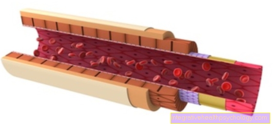
The patient initially receives a positive contrast agent, the not absorbed by the intestine and therefore remains in the lumen. The intestine is then filled with a negative contrast medium, which ensures that the positive contrast medium is pressed against the intestinal walls and pushed through the entire intestine. As a result, the entire intestinal walls are wetted with the positive contrast agent administered first. This causes an increase in the signal intensity on the intestinal walls, which thereby glow brightly in the imaging. The negative contrast agent that is in the lumen of the intestine however, reduces the signal intensity. This appears the intestinal lumen darker on imaging and can be well demarcated against the intestinal walls.
This contrasting is called Double contrast and allows the doctor to assess the intestinal walls. The thickness of the intestinal walls, their folds and constrictions (Stenoses) of the intestine, abscesses, fistulas and tumorous masses are made visible.
Especially with the Diagnosis of chronic inflammatory bowel diseases (Crohn's disease, Ulcerative colitis), the Sellink examination method using the double-contrast technique plays a major role.


.jpg)