How does seeing work?
Synonyms in a broader sense
Medical: visual perception, visualization
Look, look
English: see, watch, look
introduction
Seeing is a very complex process that has not yet been fully elucidated in detail. Light is passed on as information in electrical form to the brain and processed accordingly.
In order to understand vision, a few terms should be known, which are briefly explained below:
-
What is light
-
What is a neuron?
-
What is the visual pathway?
-
What are the optical centers of vision?

Figure eyeball
- Optic nerve (optic nerve)
- Cornea
- lens
- anterior chamber
- Ciliary muscle
- Vitreous
- Retina
What is sight
Seeing with the eyes is the visual perception of light and transmission to the visual centers in the brain (CNS).
This is followed by the assessment of the visual impressions and a possible subsequent reaction to it.
The light triggers a chemical reaction in the eye on the retina, which creates a specific electrical impulse that is passed on via nerve pathways to higher, so-called optical brain centers. On the way there, namely already in the retina, the electrical stimulus is processed and prepared for the higher centers in such a way that they can deal with the information provided accordingly.
In addition, one must also include the psychological consequences that result from what is seen. After the information in the visual cortex of the brain has become conscious, an analysis and interpretation take place. A fictitious model is created to represent the visual impression, with the help of which the concentration is directed to specific details of what is seen. The interpretation depends heavily on the individual development of the viewer. Experiences and memories involuntarily influence this process, so that each person creates his "own image" from a visual perception.
What is light
The light that we perceive is electromagnetic radiation with a wavelength in the range of 380 - 780 nanometers (nm). The different wavelengths of light in this spectrum determine the color. For example, the color red is in a wavelength range of 650 - 750 nm, green in the range of 490 - 575 nm and blue at 420 - 490 nm.
Taking a closer look, light can also be broken down into tiny particles, so-called photons. These are the smallest units of light that can create a stimulus for the eye. In order for the stimulus to be noticeable, an incredible number of these photons have to trigger a stimulus in the eye.
What is a neuron?
A Neuron generally denotes a Nerve cell.
Nerve cells can take on very different functions. ly, however, they are receptive to information in the form of electrical impulses, which can change depending on the type of nerve cell and via cell processes (Axons, Synapses) then pass it on to one or, much more often, several other nerve cells.
Illustration of nerve endings (synapse)
- Nerve endings (dentrite)
- Messenger substances, e.g. dopamine
- other nerve ending (axon)

What is the visual pathway
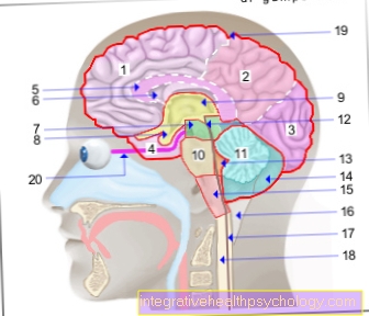
As Visual pathway the connection of eye and brain denoted by numerous nerve processes. Starting at the eye, it begins with the retina and sits in the Optic nerve into the brain. in the Corpus geniculatum laterale, near the thalamus (both important brain structures) there is then a switchover to visual radiation. This then radiates into the rear lobes (occipital lobes) of the brain, where the visual centers are located.
What are the optical centers of vision?
Optical centers of vision are areas in the brain that mainly process information that comes from the eye and initiate appropriate reactions.
This mainly includes the Visual cortexwhich is located in the back of the brain. It can be divided into a primary and a secondary visual cortex. Here what is seen is first consciously perceived, then interpreted and classified.
There are also smaller visual centers in the brain stem that are responsible for eye movements and eye reflexes. They are not only important for healthy vision, they also play an important role in examinations, for example to determine which part of the brain or the visual pathway is damaged.
Visual perception in the retina
In order for us to see, the light has to reach the retina at the back of the eye. It first falls through the cornea, pupil and lens, then crosses the vitreous humor behind the lens and must first penetrate the entire retina itself before it gets to the places where it can trigger an effect for the first time.
The cornea and lens are part of the (optical) refractive apparatus, which ensures that the light is refracted correctly and that the entire image is precisely reproduced on the retina. Otherwise the objects would not be perceived clearly. This is the case, for example, with nearsightedness or farsightedness.
The pupil is an important protective device that regulates the incidence of light by expanding or contracting. There are also drugs that override this protective function. This is necessary after operations, for example, when the pupil needs to be immobilized for some time so that the healing process can be promoted better.
Once the light has penetrated the retina, it hits cells called rods and cones. These cells are sensitive to light.
They have receptors (“light sensors”) that are bound to a protein, more precisely to a G protein, the so-called transducin. This special G-protein is bound to another molecule called rhodopsin.
It consists of a vitamin A part and a protein part, the so-called opsin. A light particle that hits such a rhodopsin changes its chemical structure by straightening a previously kinked chain of carbon atoms.
This simple change in the chemical structure of the rhodopsin now makes it possible to interact with the transducin. This also changes the structure of the receptor in such a way that an enzyme cascade is activated and signal amplification occurs.
In the eye, this leads to an increased negative electrical charge on the cell membrane (hyperpolarization), which is transmitted as an electrical signal (transmission of vision).
The Uvula cells are located at the point of sharpest vision, also called the yellow point (macula lutea) or in specialist circles called fovea centralis.
There are 3 types of cones, which differ in that they react to light of a very specific wavelength range. There are the blue, green and red receptors.
This covers the color range that is visible to us. The other colors mainly result from the simultaneous, but differently strong, activation of these three cell types. Genetic deviations in the blueprint of these receptors can lead to the various color blindnesses.
The Rod cells is found predominantly in the border area (periphery) around the fovea centralis. Rods do not have receptors for different color ranges. But they are much more sensitive to light than the cones. Their tasks are to enhance contrast and see in the dark (night vision) or in low light (twilight vision).
Night vision
You can test this yourself by trying to fix a small and just recognizable star at night when the sky is clear. You will find that the star is easier to see if you look past it lightly
Transmission of stimuli in the retina
In the Retina 4 different cell types are mainly responsible for the transmission of the light stimulus.
The signal is not only transmitted vertically (from the outer retinal layers towards the inner retinal layers), but also horizontally. The horizontal and amacrine cells are responsible for horizontal transmission, and the bipolar cells for vertical transmission. The cells influence each other and thereby change the original signal that was initiated by the cones and rods.
The ganglion cells are located in the innermost layer of nerve cells in the retina. The cell processes of the ganglia then pull to the blind spot, where they become Optic nerve (optic nerve) focus and leave the eye to enter the brain.
At the blind spot (one on each eye), i.e. at the beginning of the optic nerve, there are understandably no cones and rods and there is also no visual perception. By the way, you can easily find your own blind spots:
Blind point
Cover one eye with your hand (since the second eye would otherwise compensate for the blind spot of the other eye), fix with the eye that is not covered an object (for example a clock on the wall) and now slowly move the free outstretched arm horizontally to the right and left at the same eye level with the thumb raised. If you have done everything correctly and have really fixed an object with your eye, you should find a point (a little to the side of the eye) where the raised thumb appears to disappear. This is the blind spot.
Further information on this:
- Blind spot
- Test your blind spot
By the way: It is not just light that can generate signals in the uvula and rods. A blow to the eye or strong rubbing triggers a corresponding electrical impulse, similar to light. Anyone who has ever rubbed their eyes will certainly have noticed the bright patterns that one then thinks they see.
Visual pathway and transmission to the brain
After the nerve processes of the ganglion cells have bundled to form the optic nerve (Nervus opticus), they pull together through a hole in the back wall of the eye socket (Canalis opticus).
Behind it, the two optic nerves meet in the optic chiasm. One part of the nerve crosses (the fibers of the medial half of the retina) to the other side, another part does not change sides (the fibers of the lateral half of the retina). This ensures that the visual impressions of a complete half of the face are switched to the other side of the brain.
Before the fibers in the corpus geniculatum laterale, part of the thalamus, are switched to another nerve cell, some optic nerve fibers branch off to deeper reflex centers in the brain stem.
The examination of the eye reflex function can therefore be very helpful if you want to locate the damaged area on the way from the eye to the brain.
Behind the corpus geniculatum laterale, it then continues via nerve cords into the primary visual cortex, which are collectively referred to as visual radiation.
This is where the visual impulses are consciously perceived for the first time. However, there is still no interpretation or assignment. The primary visual cortex is arranged retinotopically. This means that a very specific area in the visual cortex corresponds to a very specific location on the retina.
The place of sharpest vision (fovea centralis) is represented on about 4/5 of the primary visual cortex. Fibers from the primary visual cortex mainly pull into the secondary visual cortex, which is laid out like a horseshoe around the primary visual cortex. This is where the interpretation of what has been perceived finally takes place. The information obtained is compared with information from other areas of the brain. Nerve fibers run from the secondary visual cortex to practically all brain regions. And so gradually an overall impression of what is seen is created, in which a lot of additional information such as the distance, movement and, above all, the assignment of what type of object it is, is incorporated.
Around the secondary visual cortex there are further visual cortex fields that are no longer ordered retinotopically and take on very specific functions. For example, there are areas that combine what is visually perceived with language, prepare and calculate the corresponding reactions of the body (e.g. "catch the ball!") Or save what is seen as a memory.
You can find more information on this topic under: Visual pathway
Way of viewing visual perception
Basically, the process of “seeing” can be viewed and described from different perspectives. The point of view described above happened from a neurobiological point of view.
Another interesting point of view is the psychological point of view. This divides the visual process into 4 levels.
The first stage (Physico-chemical level) and second step (Physical level) describe more or less similar the visual perception in a neurobiological context.
The physical-chemical level relates more to the individual processes and reactions that take place in a cell and the physical level summarizes these events in their entirety and considers the course, interaction and result of all individual processes.
The third (psychic level) tries to describe the perceptual event. This is not that easy to the extent that one cannot grasp what is visually experienced, neither energetically nor spatially.
In other words, the brain “invents” a new idea. An idea based on what is visually perceived that only exists in the consciousness of the person who has visually experienced. To date, it has not been possible to explain such perceptual experiences with purely physical processes, such as electrical brain waves.
From a neurobiological point of view, however, one can assume that a large part of the perceptual experience takes place in the primary visual cortex. On the fourth stage then the cognitive processing of the perception takes place. The simplest form of this is knowledge. This is an important difference to perception, because this is where an initial assignment takes place.
Using an example, the processing of what is perceived will be clarified at this level:
Assume that a person is looking at a picture. Now that the image has become conscious, cognitive processing begins. The cognitive processing can be divided into three work steps. First there is a global evaluation.
The image is analyzed and objects are categorized (e.g. 2 people in the foreground, a field in the background).
This initially creates an overall impression. At the same time, this is also a learning process. Because through the visual experience, experiences are gained and the things seen are assigned priorities, which are based on appropriate criteria (e.g. importance, relevance for problem solving, etc.).
In the case of a new, similar visual perception, this information can then be used and processing can take place much more quickly. Then it goes to the detailed evaluation. After a renewed and closer inspection and scanning of the objects in the picture, the person proceeds to analyze the salient objects (for example recognizing the person (couple), action (holding each other)).
The last step is the elaborative evaluation. A so-called mental model is developed, similar to an idea, but into which information from other areas of the brain now also flow, for example memories of the people recognized in the image.
Since, in addition to the visual perception system, many other systems exert their influence on such a mental model, the evaluation must be viewed as very individual.
Each person will evaluate the image in a different way on the basis of experience and learning processes and accordingly concentrate on certain details and suppress others.
An interesting aspect in this context is modern art:
Imagine a simple white picture with only a red blob of paint. It can be assumed that the splash of color will be the only detail that will attract the attention of all viewers, regardless of experience or learning processes.
The interpretation, on the other hand, is left free. And when it comes to the question of whether this is a matter of higher art, there is certainly no general answer that would apply to all viewers.
Differences to the animal world
The way of seeing described above relates to the visual perception of people.
Neurobiologically, this form hardly differs from perception in vertebrates and molluscs.
Insects and crabs, on the other hand, have so-called compound eyes. These consist of approx. 5000 individual eyes (ommatids), each with their own sensory cells.
This means that the viewing angle is much larger, but on the other hand the resolution of the image is much lower than that of the human eye.
Therefore, flying insects also have to fly much closer to objects they see (e.g. cake on the table) in order to recognize and classify them.
Color perception is also different. Bees can perceive ultraviolet light, but not red light. Rattlesnakes and pit vipers have a heat ray eye (pit organ) with which they see infrared light (heat radiation) like body heat. This is likely to be the case with night butterflies as well.
Related topics
You will also find a lot of information on related topics:
- Ophthalmology
- eye
- optical illusion
- Astigmatism
- Astigmatism baby
- Corneal inflammation
- myopia
- Visual pathway
- Lasik
- Adie syndrome
- Astuteness
- Inflammation of the optic nerve
A list of all ophthalmic topics that we have already published can be found at:
-
Ophthalmology A-Z


.jpg)