Adductors
introduction
The adductors are used to bring a part of the body closer to the body (adduction = leading, Latin adducere = leading, pulling). The adductors belong to the group of skeletal muscles.
Their antagonists are the abductors, which pull a part of the body away from the trunk. The adductors of the thigh are divided into three layers.
The superficial, middle and deep adductor groups.
All adductors are innervated by the obturator nerve, with the exception of the pectineus and adductor magnus muscles, which also receive fibers from the femoral nerve and the sciatic nerve.
In addition to the adductors of the thigh, there is also an adductor on the foot and one on the hand.
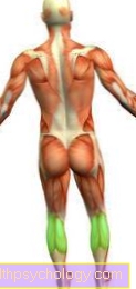
Illustration of the adductors

Adductors
- External hip muscle -
Obturator muscle
extermus - Little dresser
Adductor minimus muscle
(covered) - Comb muscle -
Pectineus muscle - Short donners -
Adductor brevis muscle - Long Dresser -
Adductor longus muscle - Great dresser
Adductor magnus muscle - Lean Muscle -
Gracilis muscle - Femur - Femur
- Kneecap - patella
- Shin - Tibia
- Femoral nerve -
Femoral nerve - Sciatic nerve -
Sciatic nerve - Hip joint -
Articulatio coxae - Pubic bone - Pubis
You can find an overview of all Dr-Gumpert images at: medical illustrations
Superficial adductor group of the thigh
The superficial group of adductors on the thigh consists of:
- Of the Pectineus muscle arises on Pecten ossis pubis (Protrusion of the Pubic bone) and continues at the Linea pectinea of Thighbone (Femur) on. He serves the Adduction of the thigh. He can also use the thigh rotate externally and bow.
- Of the Adductor longus muscle has its origin in the upper part of the pubic bone (Ramus superior of the os pubis) and pulls to the middle part of the Linea aspera of the femur. This is centered on the back of the thigh bone. This muscle also serves the Adduction and bends also the thigh in the hip joint.
- Of the Gracilis muscle arises in the lower part of the pubic bone ( Ramus inferior of the pubic bone) and at the Symphysis. He pulls up to Shin bone (Tibia) and sits there below the tibial head together with the Sartorius muscle and the Semitendinosus muscle on. Since the gracilis muscle extends across both the hip and the Knee joint pulls away, he's the only one two-jointed muscle of the entire adductor group. It causes both the diffraction as well as the Adduction, in the knee joint he is at the Internal rotation and diffraction involved.
Appointment with ?

I would be happy to advise you!
Who am I?
My name is I am a specialist in orthopedics and the founder of .
Various television programs and print media report regularly about my work. On HR television you can see me every 6 weeks live on "Hallo Hessen".
But now enough is indicated ;-)
In order to be able to treat successfully in orthopedics, a thorough examination, diagnosis and a medical history are required.
In our very economic world in particular, there is too little time to thoroughly grasp the complex diseases of orthopedics and thus initiate targeted treatment.
I don't want to join the ranks of "quick knife pullers".
The aim of any treatment is treatment without surgery.
Which therapy achieves the best results in the long term can only be determined after looking at all of the information (Examination, X-ray, ultrasound, MRI, etc.) be assessed.
You will find me:
- - orthopedic surgeons
14
You can make an appointment here.
Unfortunately, it is currently only possible to make an appointment with private health insurers. I hope for your understanding!
For more information about myself, see - Orthopedists.
Middle adductor group of the thigh
To middle adductor group only belongs to Adductor brevis muscle.
Like the gracilis muscle, this arises from the lower part of the pubic bone (Ramus inferior of the pubic bone) and continues on the middle part of the Linea aspera of the thigh bone at (Labium mediale of the linea aspera of the femur).
This also serves the Adduction and contributes a small part diffraction and External rotation in the hip joint.
Deep adductors of the thigh
The deep adductor group is made up of the glarge (magnus) and small (minimus) adductor muscles together:
- Of the Adductor magnus muscle arises from the ischial tuberosity (Sciatic tuberosity) and the smaller part of the ischium (Ramus ossis ischii).
He puts on the middle part of the Linea aspera at the back of the thigh bone (Labrum mediale of the linea aspera of the femur) on. Another large part of this muscle finds its insertion on the Medial epicondyle of the thigh.
This is considered the strongest adductor of the entire adductor group. In addition he stretches the thigh in the hip joint. In addition, the (proximal) Muscle fibers rotate the thigh outwards, the (distal) Parts rotate the thigh inwards. - Of the Adductor minimus muscle is considered one Splitting off of the great adductor muscle. It has the same origin and insertion as the adductor magnus muscle. Its function is to Adduction and External rotation of the thigh in the hip joint.
Adductors of the foot
Of the Adductor hallucis muscle consists of two muscle heads that have a different origin.
The Caput transversum arises at the joint capsule of the 3rd to 5th metatarsophalangeal joint, the Caput obliquum arises at the cuboid (Os cuboideum), on the outer sphenoid bone (Os cuneiform laterale) and on 2-4. Metatarsal bones.
The common approach can be found at the base of the big toe.
This will make the Big toe adducted, so brought up to the second toe. This muscle is innervated by the Lateral plantar nerve.
Adductors of the hand
Of the Adductor pollicis muscle has a similar structure to the adductor of the foot.
This muscle also consists of two muscle heads.
The Caput obliquum arises from the head leg (Os capitatum), the Caput transversum finds its origin in the 3rd metacarpal bone (Os metacarpal III).
Both heads attach to the medial side of the sesamoid bone.
This causes the muscle to Adduction of the thumb. Furthermore, he is for the Opposition movement in the D.saddle joint essential. The thumb is moved to the palm of the hand.