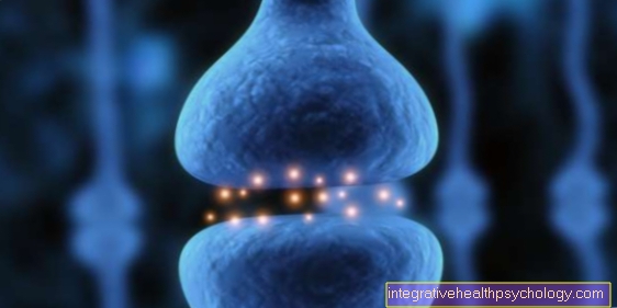
Action potential
Synonyms
Nerve impulse, excitation potential, spike, excitation wave, action potential, electrical excitation
definition
The action potential is a brief change in a cell's membrane potential from its resting potential. It is used to transmit electrical excitation and is therefore elementary for the transmission of stimuli.

physiology
In order to understand the action potential, one must first look at the Resting potential become aware of a cell. Every excitable cell in the resting state has one. It is created by the Difference in charge between the inside and outside of the Cell membrane and it depends on the respective cell how high it is. As a rule, the values fluctuate between -50 mV and -100 mV. Most nerve cells have a resting potential of -70mV, which means that in the resting state the inside of the cell membrane is negatively charged compared to the outside of the cell membrane. We will now look at the development of an action potential using a nerve cell. Here action potentials cause a quick one Excitation conduction in the body over long distances.
Starting position
The cell has a resting membrane potential, which is maintained by the sodium-potassium pump.
Initiation phase
An excitation, triggered by a stimulus, reaches the cell. The inside of the cell becomes more positive due to the inflowing sodium ions. If a certain threshold value is exceeded (in the case of nerve cells approx. - 50mV) an action potential is triggered. This works according to the “all or nothing principle”. That means there is no such thing as “a little potential for action”, either it arises or it doesn't. The shape of the action potential is always uniform after the threshold value is exceeded, regardless of the strength of the stimulus.
Depolarization
If the threshold value is exceeded, many sodium channels on the cell membrane open in one fell swoop and many sodium ions flow into the interior of the cell from the outside at once. The cell becomes positive inside with up to approx. +20 to + 30 mV. This event is also known as the "spread" or "overshoot".
Repolarization
After the maximum of the spread is reached, the sodium channels begin to close again. For this, potassium channels open, with which positively charged potassium ions flow out of the cell and the inside of the cell becomes more negative again.
Hyperpolarization
As a result of the repolarization, the resting potential is usually not reached at first and can reach values of up to -90 mV, for example in the case of a nerve cell with a resting potential of -70 mV. This is also called hyperpolarizing postpotential. It arises from the fact that the potassium channels close more slowly and thus more positively charged potassium ions flow out of the cell.
The original ratio is then restored by the sodium-potassium pump, which uses energy to transport three sodium ions out of the cell and, in return, transports two potassium ions into the cell.
The so-called refractory phase is also important for the action potential. It arises from the fact that the sodium channels are inactive for a short time after the action potential has been triggered. Thus no further action potential can be triggered during the “absolute refractory period” and a further action potential can only be triggered to a limited extent during the “relative refractory period”.
An action potential lasts about 1-2 milliseconds in nerve cells. In a heart muscle cell it can even be several hundred milliseconds.
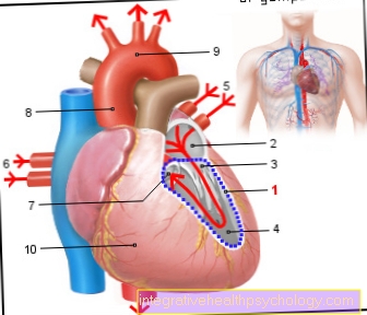
Action potential at heart
The basis of electrical stimulation in the heart is the so-called action potential.It represents the biologically time-limited change in an electrical voltage across the cell membrane, which ends in a muscle action, in this case the heartbeat. With a duration of around 200 to 400 milliseconds depending on the respective heart rate, i.e. the number of heartbeats per minute, that is Action potential on the heart longer than that of a skeletal muscle or nerve cell. This protects the heart from overexcitation.
Starting from a certain resting potential, a basic voltage of around minus 90 millivolts, which is applied to the membranes of the cells, the action potential runs through the heart four phases of arousal formation. Different ion channels work together to change the electrical voltage on the outside of the cells. These are mostly transport proteins that are located in the skin of the cells and transport various very small charged particles across their membrane. This will make the electrical voltage on the cell changes and thus formed the action potential on the heart.
In the first phase, the so-called Depolarization phase, the ability to transport positively charged sodium particles increases. These now flow into the interior of the cells and lead to one Increase in tension from about minus 90 millivolts to plus 30 millivolts.
By shifting the electrical charge into the positive range, they become specific Calcium channels at heart open. So it comes to one Influx of calcium particles into the heart cells. These second phase represents the long lasting that is typical of the heart Plateau phase This is where the excitement is carried and prevents, among other things, the entry of additional superfluous action potentials. It ensures the controlled pumping capacity of the heart and protects against cardiac arrhythmias.
In the third phase, the Repolarization phase, the electrical voltage slowly returns in the direction of the rest potential of minus 90 millivolts. Due to an energy-consuming process, contrary to the concentration gradient above the cell, the inflowing becomes active Sodium particles back outside and emanated Potassium parts back into the cell transported. And this until the original resting potential has leveled off again. The cell is now ready for a new action potential.
Action potential at the sinus node
The excitation origin of the action potential at the heart lies in the so-called Sinus node. This is located in the right auricle near the confluence of the superior vena cava, which transports the blood from the upper body circulation to the heart.
The sinus node consists of modified muscle cellsthat create the action potentials necessary for arousal. They thus form the natural one Pacemaker of our heart. These are quickly excitable cells with a natural frequency of around 60 to 80 beats per minute. This natural frequency can be registered in the form of the pulse.
From there, the resulting action potential takes its course via certain anatomical structures in order to lead to a contraction, a heartbeat, in the working muscles of the heart. The number of beats per minute can be adapted to the load on the person. The Sympathetic, an autonomous nervous system that is especially important when increasing burden is activated, leads to an increase in the incoming action potentials.
Will the opposite, the so-called Parasympathetic nervous system activated, especially in Rest periods of the body plays a role, the number of action potentials towards the heart is throttled. The heartbeat slows down. Also Medication and the body's own Hormones, like adrenaline, affect this system.


-whrend-der-schwangerschaft.jpg)