Anatomy Lexicon
Explanation
Why a Anatomy Lexicon?
In order to be able to understand many diseases better, it is important to know the function “in the healthy”.
In this anatomy dictionary you will find many anatomical terms such as bone, Joints and Muscles described.
There is a link to the corresponding clinical pictures.
definition

anatomy denotes the Teaching the structure of organisms. As a large field of morphology it is very important in everyday medical practice. In human and veterinary medicine (veterinary medicine), for example, anatomy describes the construction of Skeleton, the Location of internal organs, the Musculature and the Course of annoy- and Vascular pathways.
When naming the individual structures of the organism, a standardized nomenclature used that on the Latin and Greek Language is justified.
Overall, the anatomy can be broken down into several Sub-areas structure.
Subsections of anatomy
The movement system
In order to be able to diagnose complaints of the musculoskeletal system, a knowledge of the anatomy is necessary.
The anatomy of the musculoskeletal system deals with the teaching of:
- Anatomy of the bones
- Tapes
- Joint anatomy
- Muscle anatomy
- Tendons
1. Anatomy of the bones
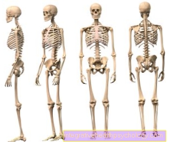
The adult human skeleton consists of more than 200 different bones, which differ greatly in shape, size and stability - depending on which tasks they have to fulfill.
skull
The skull consists of many different bones that are firmly fused with one another in adult humans.
It is further subdivided into brain skull (lat .: Neurocranium) and facial skull (lat .: Viscerocranium).
For more information, see: skull
Humerus
The upper arm bone is also known medically as the humerus. The humerus forms the shoulder joint with the shoulder blade and the elbow joint with the ulna and radius.
Read more on the topic: Humerus
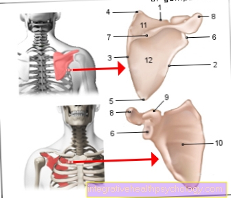
shoulder blade
The shoulder blade (lat .: Scapula) is a flat, roughly triangular bone and the connection between the upper arm and the trunk.
The shoulder height, the outer area of the shoulder blade, forms together with the collarbone (lat .: Clavicle) and the humerus, the shoulder joint.
For more information, see: shoulder blade
Cubit
The ulna is also medically called Ulna designated. It forms with the spoke (radius), the bones of the forearm.
For more information, see: Cubit
spoke
The spoke (lat .: radius) forms with the cubit (lat .: Ulna), the bones of the forearm.
For more information, see: Spoke
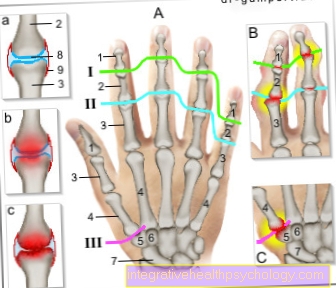
Carpal
The wrist is made up of 8 small bones that together form the bony skeleton of the hand. They are located in two different rows, the first of which, together with the spoke, forms the wrist.
For more information, see: Carpal
Collarbone (Clavicle)
The collarbone (lat .: Clavicle) is an approximately 12-15 cm long, S-shaped bent bone.
It belongs to the shoulder girdle and connects the breastbone (sternum) with shoulder height (lat .: Acromion), part of the shoulder blade (lat .: Scapula).
For more information, see: Collarbone
Rib cage
The chest (lat .: thorax) bony encloses the lungs and the heart.
It is formed by the ribs, the sternum and the thoracic spine.
In addition to this protective function, it plays an essential role in breathing.
For more information, see: Rib cage
Sternum
On the sternum (lat .: sternum) the ribs end (lat .: costae) on the front of the chest (lat .: thorax).
For more information, see: Sternum
Ribs
Humans have 12 pairs of ribs (lat .: costae), which are all connected to our thoracic spine and determine the shape of the rib cage.
They protect the organs of the chest and are an important part of the respiratory system.
For more information, see: Ribs
Pelvic bones
The bony pelvis consists of different bones: the two hip bones (Os coxae), the tailbone (Os coccygis) and the sacrum (Sacrum). It articulately connects the spine and lower extremity.
The bony structure of the pelvis differs between the sexes due to the anatomical requirements for the birth of a child.
For more information, see: Pelvic bones
Thigh bone
The thigh bone (lat .: Femur) represents the only bone of the thigh. It transfers the force from the pelvis to the knee joint.
For more information, see: Thighbones
Kneecap
The kneecap (lat .: patella) belongs to the knee joint. Their task is to redirect the force of the thigh muscles to the shin (lat .: Tibia).
For more information, see: Kneecap
Shin
The shin (lat .: Tibia) transfers almost 100% of the force from the knee to the upper ankle.
For more information, see: Shin
Fibula
The fibula and the tibia form the two bones of the lower leg.
The fibula only plays a subordinate role in the knee joint. At the ankle, it forms the outer ankle.
For more information, see: fibula
Foot bones
Similar to the hand, the foot also consists of several small bones that are connected to one another with ligaments.
The largest bones of the foot are the ankle bone, which together with the tibia and fibula form the upper ankle joint, and the heel bone, an important part of the lower ankle joint.
For more information, see: Anatomy of the foot
Spine
The spine is divided into the cervical spine (HWS), the thoracic spine (BWS) and the lumbar spine (LWS). It is extremely important for the statics of the human body.
Overall, the spine describes an S-shaped curvature in a healthy person. This special shape is used to cushion impacts.
The spine is made up of alternating bony vertebral bodies and the intervertebral discs. There are small joints between the vertebrae that allow the body to tilt forwards and sideways
For more information, see: Spine anatomy
- Cervical spine
The cervical spine (cervical spine), the uppermost section of the spine, consists of 7 vertebrae.
The top two, carriers (lat .: Atlas) and lathe operator (lat .: Axis) form the head joint, which enables the head to be turned and tilted.
For more information, see: Cervical spine
- Thoracic spine
The ribs, which extend from the back to the sternum, attach to the twelve vertebrae of the thoracic spine (thoracic spine) and thus determine the bony shape of the rib cage.
As a result, the thoracic spine is less mobile than the other sections of the spine.
For more information, see: Thoracic spine
- Lumbar spine
The lumbar spine, which consists of 5 vertebral bodies, bears the main weight of the body.
It connects the thoracic spine and the pelvic area, where it connects to the sacrum (lat .: Sacrum) is in communication.
For more information, see: Lumbar spine
2. Ribbons
Ligaments are made up like tendons connective tissue fibers. However, they do not connect muscles and bones, but movable parts of the bony skeleton.
They are much more rigid than tendons, and serve the Stabilization of bones and joints. In this way, they specify the possible range of motion of a joint and keep highly stressed areas in shape.
This is particularly pronounced, for example, on Ankle joint.
3. Anatomy of the joints
The joints are an important part of the musculoskeletal system: only through them is it possible for bones not to be rigidly fixed, but also to be mobile against each other.
This will give you an overview of the most important joints in the human body.
Shoulder joint
The shoulder joint connects the shoulder blade and the upper arm.
In contrast to other joints, it is only held by a few ligaments, which enables a large number of movements.
It is secured by the strongly pronounced Shoulder muscles, especially the so-called Rotator cuff is relevant.
For more information, see: Shoulder joint
Elbow joint
The elbow joint consists of three partial joints which, in their entirety, connect the lower and upper arm bones.
It enables both extension and flexion as well as rotation of the forearm.
For more information, see: Elbow joint
wrist
The wrist is of spoke (lat .: radius), Cubit (lat .: Ulna) and the first row of carpal bones (esp. scaphoid and lunar bone).
Further information on this topic is available at: wrist
hip joint
The hip joint connects the pelvis and thighbones.
It is held in place by very strong ligaments, as it must also remain stable under the entire body weight.
It enables extension, flexion, rotation and spreading movements of the leg.
For more information, see: hip joint
Knee joint
The knee is the largest joint in the human body and has a very complex structure. The two joint partners are the Thighbones (Femur) and the Shin (Tibia). Also the Kneecap (patella) is involved in the knee joint.
They run inside the knee joint front and the posterior cruciate ligament. Together with other ligaments, they stabilize the knee so that the upper and lower legs cannot move against each other.
The menisci are also an important part of the knee
For more information, see: Knee joint
Ankle joint
The ankle joint connects the foot and lower leg. Strictly speaking, there are not one but two joints:
- Upper ankle
The upper ankle is made up of three bones, the shinbone (tibia), the fibula (fibula) and finally the ankle bone (talus).
For more information, see: Upper ankle
- Lower ankle
The lower ankle connects Ankle bone, Calcaneus and Scaphoid together.
it enables pronation (turning outward) and supination (turning inward) of the foot.
For more information, see: Lower ankle
4. Muscle anatomy
Our body has around 650 muscles without which humans would not be able to perform any movements. An upright posture is also only possible in this way.
This will give you an overview of the most important muscle groups in the human body.
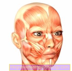
Neck muscles
The so-called short neck muscles pull from the Cervical spine to the head.
They allow the head to be tilted forwards, backwards and sideways.
For more information, see: Neck muscles
Shoulder muscles
The muscles of the shoulder arise shoulder blade, Rib cage or Spine, and pulls to the upper arm.
It consists of a large number of muscles, which are further subdivided depending on their location and function.
The so-called Rotator cuffthat surrounds the arm like a cuff is also part of the shoulder muscles. Since the shoulder joint is only held by a few ligaments, it is very important for its stabilization.
For more information, see: Shoulder muscles and Rotator cuff
Arm muscles
The muscles of the Upper arm serves the Flexion and extension in the elbow joint.
it consists of biceps, Triceps, Arm flexors, Upper arm radial muscle and Cartilaginous muscle (lat .: Anconeus muscle).
Of these muscles, the triceps is the only extensor of the elbow joint.
The muscles of the Forearm not only moves the hand and elbow, but also the fingers.
It is further divided according to its position (front or back of the arm) and according to its function (flexor and extensor).
For more information, see: Arm muscles
Chest muscles
The chest muscles consist of the Pectoralis major (lat .: M. pectoralis major) and the Small pectoral muscle (lat .: M. pectoralis minor).
It enables the arm to be guided towards the body (Adduction), swinging the arm forward (Anteversion) as well as the Internal rotation of the arm.
For more information, see: Chest muscles
Abdominal muscles
The abdominal muscles are made up of the straight abdominal muscle and the obliques educated.
They allow the body to bend and tilt sideways.
For more information, see: Abdominal muscles
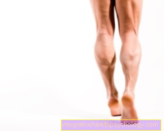
Leg muscles
The muscles of the leg are divided into thigh and lower leg muscles.
The Thigh muscles pulls from the pelvis and hip area to the thigh.
According to their function, the individual muscles are counted as extensors or flexors.
ly they allow movement in the hip, but some muscles also act on the knee joint.
In addition, the thigh muscles stabilize the hip joint when standing.
The Lower leg muscles enables movements in the ankle. It is divided into two subgroups according to function and location: those lying on the front of the leg Straightener and those behind Flexor.
For more information, see: Thigh muscles and Lower leg muscles
Back muscles
The long back muscles (lat .: M. erector spinae) acts as an opponent of the straight line Abdominal muscles and thus takes over the stretching of the Spine.
It is also very important for maintaining an upright posture.
For more information, see: Back muscles
5. tendons
Tendons are made of fibers connective tissue, the the Train transmission serve between muscle and bone.
They represent the end of the muscles with which they attach to the bone.
Additionally, there are also tendons that run between muscle bellies as well flat tendon plates (Aponeuroses) such as on the palm of the hand.
For more information, see: Tendons
Tendon sheaths
A tendon sheath is a tubular structure that is a tendon as a Guide channel surrounds.
This protects the tendon from mechanical injuries protected.
Tendon sheaths occur in places where tendons have to be guided around or through other anatomical structures, such as protruding bones, ligaments or joints.
For more information, see: Tendon sheath
Biceps tendon
The Biceps muscle (Biceps brachii muscle) has two sinewy Origins.
The des long head arises at the upper edge of the pan Shoulder joint, the des short head on the raven-beak process, a bone process on the shoulder blade.
The common one approach of both muscle heads lies on a roughened part of the spoke, the Radial tuberosity.
For more information, see: Biceps tendon
Achilles tendon
The approximately 15 to 20 cm long Achilles tendon (lat .: tendo calcanei) is the starting point of the three-headed calf muscle (lat .: Triceps surae muscle).
All three muscle heads unite in their course, creating the Achilles tendon. They work together on that Calcaneus at.
For more information, see: Achilles tendon
Patellar tendon
The patellar tendon pulls away from the Kneecap to a rough place of the Tibia, the so-called Ttibial excess.
Strictly speaking, it is not a separate tendon, but an extension of the tendon of the quadruple thigh muscle (lat .: Quadriceps femoris muscle).
For more information, see: Patellar tendon
Organ systems
Internal organs anatomy
The anatomy of the internal organs includes different organ systems. In the following you will get an overview of the internal organs:
- Respiratory tract
- Cardiovascular system
- Digestive system
- Genital organs
- Urinary tract
- Glands
1. Respiratory tract
The Respiratory tract is needed to supply the body with oxygen.
It consists of the windpipe (lat .: Trachea), the Larynx (lat .: larynx) and the various sections of the lung (lat .: Pulmo).
Further information on this topic can be found at: Respiratory tract
Larynx
The Larynx (lat .: larynx) connects the throat (lat .: Pharynx) with the windpipe (lat .: Trachea).
It mainly serves the breathing and the Voice training.
He is also involved in the swallowing process and prevents as Valve the penetration of food and drink into the deeper airways.
For more information, see: Larynx
windpipe
The trachea is a 10-12 cm long, elastic tube that connects the larynx with the lungs.
With reference to the Spine the trachea begins at the level of the 6th / 7th. Cervical vertebra and ends at the level of the 4th thoracic vertebra.
There it divides into the left and right bronchus which then pull into the lung tissue.
For more information, see: windpipe
Bronchi
The bronchi are the airways within the lungs. They are divided into an air-conducting and a respiratory part, in which the gas exchange takes place.
The bronchi begin at the branching off of the windpipe at the level of the 4th thoracic vertebra with the two large ones bronchi.
These then divide into the two Lungs and branch up to the tips of the lungs.
In this way the bronchi get smaller and smaller until they are as Alveoli (Alveoli) at which the actual gas exchange takes place.
For more information, see: Bronchi
lung
The lung (lat .: Pulmo) is the body's organ that is responsible for an adequate intake and supply of oxygen.
It consists of two spatially and functionally independent lungs and with these surrounds the heart. The two organs lie in common Rib cage, protected by the Ribs.
The lungs have no shape of their own but are shaped in their relief by the surrounding structures (diaphragm below, heart in the middle, outside the ribs, above the trachea and esophagus).
For more information, see: lung and Breathing process
Alveoli
The around 400 million alveoli (lat .: Alveoli) are the smallest unit of the lungs.
This is where the main part of the gas exchange takes place: the oxygen from the inhaled air is absorbed into the bloodstream through the wall of the alveoli.
For more information, see: Alveoli
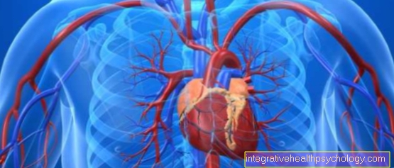
2. Cardiovascular system
The Heart-The circulatory system is used to supply the body with oxygen and nutrients via the Arteries, as well as the Removal of "waste products" of the metabolism via the Veins.
It is divided into the small and large circulation according to its function.
- The small cycle leads the oxygen-poor blood via the veins to the right heart, from where it is pumped into the lungs for gas exchange.
- The great cycle distributes the oxygen-rich blood, which comes directly from the lungs, to the whole body via the left heart in order to supply it.
Further information on this topic is available at: Cardiovascular system
The heart
The heart is a large muscular organ that that blood pumps through the body.
Functionally, the heart is made up of two chambers of the heart, each with a Atrium are connected. It lies in the middle skin (Mediastinum) between the two lungs and is protected from the outside by the bony chest (thorax). It is surrounded by the pericardium (lat .: Pericardium).
They run on the outside of the heart Coronary arteriesthat supply blood to the heart itself.
Also has the heart own veinsthat transport the oxygen-poor blood from the heart muscle and direct it to the right atrium.
For more information, see: heart
Atria
The heart has two atria, the right and the left atrium.
The atria are the respective ventricle (Ventricle) upstream.
- Right atrial
The right atrium is part of the small circulation (also Pulmonary circulation called):
The venous blood gets out of the body via the Vena cava (upper and lower Vena cava) into the forecourt, passes the right wing valve (Tricuspid valve) and flows into the right ventricle.
From here the blood is pumped to the lungs, where it is reloaded with oxygen.
For more information, see: Right atrial
- Left atrium
The left atrium is part of the great circulation (also Body circulation called):
The blood, which was previously saturated with oxygen in the lungs, passes through the Pulmonary veins into the forecourt, and via the left leaflet valve (Mitral valve) in the left ventricle.
Here it is about the artery (aorta) pumped into the body periphery.
For more information, see: Left atrium
Chambers of the heart
As heart chambers (lat .: Ventricle) are the names of the two large heart cavities.
- Right ventricle
The right ventricle is part of the pulmonary circulation and the right atrium (Atrium dextrum) downstream.
She pumps that deoxygenated blood into the pulmonary arteries, where it is again saturated with oxygen, and then enters the circulatory system via the left heart.
For more information, see: Right ventricle
- Left ventricle
The left ventricle is part of the Body circulation the left atrium (Atrium sinistrum) downstream.
The fresh from the lungs oxygenated blood is drawn from the left ventricle into the main artery (the aorta) is pumped so that it can supply all important structures with oxygen.
For more information, see: Left ventricle
Pericardium
The Pericardium (lat .: Pericardium) is a connective tissue covering that encloses the heart.
On the one hand it serves to protect the heart from external influences, but on the other hand it also prevents excessive expansion of the heart.
For more information, see: Pericardium
Blood vessels
A Blood vessel is a Hollow organ with a certain cell structure.
Blood flows in almost every area of our body, with the exception of the cornea eye (Cornea), the Enamel, hair and Nails.
For more information, see: Blood vessels
Different types of blood vessels are distinguished depending on their thickness and function:
- Arteries
An artery is a blood vessel that draws blood from the heart leads away. So you transport that oxygen-rich Blood to various organs and tissues.
The only exception is the pulmonary artery: this carries the deoxygenated blood from the right ventricle to the lungs, where it is again enriched with oxygen.
The largest artery in the human body is the artery (aorta). Depending on the body size, it has a diameter of up to three centimeters.
The smallest arteries will Arterioles called: They are no more than a tenth of a millimeter thick.
For more information, see: Arteries
- Veins
As vein is called a Blood vesselwhat blood to the heart leads there. So oxygen-poor blood always flows through the veins
The only exception is again formed by the pulmonary veins: These transport the freshly oxygenated blood to the heart.
Compared to Arteries veins have a different structure and different functions: they have and have a much thinner muscle wall Venous valvesthat prevent the blood from flowing back.
The largest veins in the body are the two so-called Vena cavathat carry the body's venous blood to the right atrium.
For more information, see: Veins
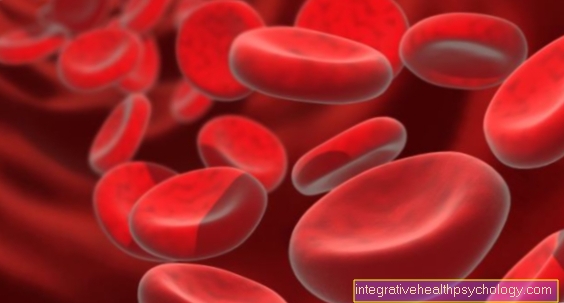
- Capillaries
Capillaries are the smallest vessels in the body. They are so small that one red blood cell (Erythrocyte) usually only fits through under its own deformation.
They represent the connection between the venous and arterial vascular system: The exchange of substances between blood and tissue takes place in them.
For more information, see: Capillaries
Heart muscle
The Heart muscle (Myocardium) consists of a special type of muscle that is found nowhere else in the body. It is particularly characterized by a type of excitation propagation and control that is unique in its form.
Only through the regular tension of the muscle, the blood is pumped from the heart into our body.
For more information, see: Heart muscle
3. Digestive system
The digestive system of humans serves that admission, digestion and Recovery of food and fluids.
It consists of a multitude of organs, which in their entirety are called Digestive tract are designated.
For more information, see: Digestive tract
esophagus
The esophagus (lat .: Esophagus) is on average 25-30 cm long in adult humans.
It is a muscle tube that runs through the oral cavity and the stomach connects and is mainly responsible for the transport of food after eating.
For more information, see: esophagus
stomach
The stomach is a muscular hollow organ between the esophagus and the Intestines lies. Its job is to mix and pre-digest the food ingested
For this purpose, the acidic gastric juice (hydrochloric acid) and enzymes are formed, which chemically break down, reduce or split some components of the food in order to then add the chyme in portions Small intestine forward.
For more information, see: stomach
Duodenum
The approximately 30cm long duodenum (Duodenum) is part of the Small intestine.
It forms the link between the stomach and the Jejunum (Jejunum).
For more information, see: Duodenum
Small intestine
The Small intestine is the section of the Digestive tractthat the stomach follows. This is divided into three sections. He starts with that Duodenum (Duodenum) followed by Jejunum (Jejunum) and Ileum (Ileum).
The small intestine is responsible for the pulp (Chyme) into its smallest components columns, as well as certain nutrients to record (to resorb).
For more information, see: Small intestine
Colon
The large intestine is the section of the digestive tract that follows the small intestine.
It is about 1.5 meters long and has the task of liquid and Minerals (Electrolytes) to be absorbed from the intestinal contents. This is how the stool is thickened.
In addition, the large intestine is colonized with bacteria that have many important functions
For more information, see: Colon
rectum
The rectum is the last section of the digestive tract. It follows the large intestine and consists of two parts:
- Rectum
The Rectum (lat .: Rectum) serves together with the anus of the Stool elimination (Bowel movement, defecation). This is where the stool is collected, and via receptors in the intestinal wall Urge to defecate triggered.
The rectum is surrounded by a variety of muscles that help control bowel movements (Continence) can be guaranteed.
For more information, see: Rectum
- nach
As nach is called the Sphincter muscle at the end of the intestinal canal. It is used to control holding back as well as passing stool from the Intestines.
For more information, see: nach
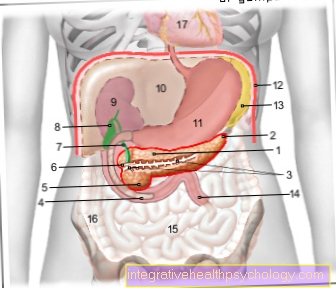
pancreas
The pancreas is a gland weighing about 80g, 14 to 18 cm long and located in the upper abdomen between Small intestine and spleen.
Due to its appearance, the entire gland in the head (lat .: Caput), Body (lat .: Corpus) and tail (lat .: Cauda) divided.
It consists of two parts: the so-called exocrine partthat produces digestive enzymes and that endocrine part, the hormones, especially insulin and Glucagon produced.
Further information on this topic can be found at: pancreas
liver
The liver is the central metabolic organ of humans, and thus also part of the digestive system.
To the Functions of the liver belongs to the food-dependent storage of sugars and fats, the breakdown and the excretion of toxins that education most blood proteins and bile, as well as numerous other tasks.
For more information, see: liver and Function of the liver
Gallbladder
The Gallbladder is a small, approximately 70 ml hollow organ that is attached to the underside of the liver lies.
The job of the gallbladder is that which is continuously formed by the liver Bile to be stored between meals and when needed for digestion in the Duodenum (Duodenum) to submit.
Further information on this topic is available at: Gallbladder
Gallstones
As Gallstones denotes deposits (concrements) in the Gallbladder (Cholecystolithiasis) or the Bile ducts (Choleangiolithiasis).
For more information, see: Gallstones
4. Sex organs
The Genital organs of humans are used for reproduction and the production of growth and gender-specific hormones.
In addition to the obvious division into female and male Sex organs become wider inner and outer Organs differentiated: The external genital organs are those that are visible from the outside, the internal genital organs are hidden in the body cavities.
Female reproductive organs
- Ovaries
The Ovaries (Ovaries) of the woman are on the right and left of the uterus (uterus) in the small pelvis.
You are the Reproductive organs of the woman:
Here they mature ova approach, and become part of the menstrual cycle in the Fallopian tubes submitted.
Important hormones for women are also produced here (especially estrogen).
For more information, see: Ovaries and Function of the ovaries
- Fallopian tubes
The Fallopian tubes connect the Ovaries with the uterus. The matured egg cell is transported and fertilized in them after ovulation.
The fallopian tube is one of the female sexual organs and is created in pairs. One fallopian tube is about on average 10 to 15 cm long. You can think of it as a hose, so to speak, that carries the Ovary with the uterus connects and thereby a matured Egg cell, which can be fertilized in the course of the fallopian tube, enables safe transport.
For more information, see: Fallopian tubes
- uterus
The uterus (lat .: uterus) belongs to the reproductive organs of women and is located in the small pelvis. It is a roughly pear-shaped organ 5 cm wide and 7 to 8 cm long.
During pregnancy, the unborn child matures in the body of the uterus
Further information on this topic is available at: uterus
- Mammary gland
The chest consists of glands (lat .: Glandula mammaria), Fat and connective tissue.
Anatomically, the breast can be divided into 10 to 12 lobes (lobi).
With the completion of the puberty the mammary gland can then take up its function:
During a pregnancy the mammary glands unfold to their full size in order to support the baby during breastfeeding Breast milk to supply.
Further information on this topic is available at: Female breast
- vagina
The vagina or vagina is one of the female genital organs and is a thin-walled one, for example 6 to 10 cm long, stretchable hose connective tissue as Musculature.
The so-called protrudes into the vagina Portio, the end of the Cervix (lat .: Cervix); its mouth is in the vaginal vestibule (Vestibulum vaginae, vestibule = Forecourt).
For more information, see: vagina
Male reproductive organs
- Testicles
The paired ones Testicles (lat .: Testis) are used for the production of sperm and hormones.
The function of the testicle is made through Pituitary gland and Hypothalamus controlled.
For more information, see: Testicles
- Epididymis
The epididymis lies above the testicle and is slightly shifted backwards (craniodorsal).
It is over an upper and a lower band (Ligamentum epididymis superior and inferior) connected to the testicle.
He is the place of Sperm maturation and Seed storage.
In addition, the epididymis is part of the executive Seminal ducts.
For more information, see: Epididymis
5. Urinary tract
The Urinary tract is, as the name suggests, responsible for the production and excretion of urine.
It consists of several parts:
In the kidney toxins and other substances requiring elimination are removed from the blood and then thickened.
About the efferent urinary tract the urine is now heading towards bladder headed to then willingly over the ureter to be eliminated.
kidney
The kidney, of which every human usually has two, is roughly bean-shaped.
Each kidney weighs approximately 120-200 g, with the right kidney generally smaller and lighter than the left.
Through the production of urine, the kidneys have an influence on many processes in the body, such as the excretion of urinary substances, long-term blood pressure control, and regulation of the water and salt balance.
For more information, see: kidney
Urinary tract
Be covered by the term "urinary tract" Renal pelvis (Pelvis renalis) and ureter summarized, which are lined by specialized tissue, the so-called urothelium.
For more information, see: Urinary tract
- Renal pelvis
The renal pelvis (lat .: Pelvis renalis) lies within the kidney and connects the kidney and ureter.
It is a space lined with mucous membrane, which is funnel-shaped to the so-called Calyx (lat .: Calices renalis) expanded.
These include the kidney papillae, which is where the urine produced in the kidney arrives.
For more information, see: Renal pelvis
- ureter
The ureter (lat .: Ureter) connects renal pelvis and bladder. It is a 30-35cm long tube made of thin muscles and mucous membrane.
It runs in the space behind the abdominal cavity (lat .: Retroperitoneum) into the pelvis, where it opens into the back wall of the urinary bladder.
For more information, see: ureter
bladder
The bladder is a muscular sac that is responsible for storing and emptying urine. The urinary bladder (Vesica urinaria) is located in the pool and when empty it is compressed by the abdominal viscera as a flaccid sack.
For more information, see: bladder
urethra
The urethra (lat .: urethra) is a muscular tube that carries urine from the bladder leads to the external urinary opening.
There are considerable differences between men and women in terms of the location and course of the urethra:
The female urethra is 3-5cm long and has a straight course.
It starts at the lower end of the bladder and passes through the pelvic floor and flows between the small ones labia minora.
The male urethra is with 20 cm significantly longer than the female.
In contrast to the female urethra, the male urethra is simultaneous Urinary and sexual tract.
The man's urethra has its origin (Ostium urethrae internum) as well as the female on the bladder neck. Then follow four anatomical sections until it ends on the outside of the glans.
For more information, see: urethra
6. glands
The Glands of the human body play one essential role in almost all bodily functions because those produced by them Hormones control and regulate a large number of tasks.
thyroid
The 20 to 25g heavy in adults thyroid belongs to the so-called endocrine organs of the body. Their main (endocrine) task is the formation of hormones that are released (secreted) into the blood.
For more information, see: thyroid
Parathyroid
The Parathyroid glands represent four lens-sized glands weighing approx. 40 mg. They lie behind the thyroid at. Usually two of them are at the top (pole) of the thyroid lobes, while the other two are at the bottom pole.
For more information, see: Parathyroid
Adrenal gland
The Adrenal glands are important hormonal glands. They lie on the kidneys like a cap, and are approx. 4 cm long, 3 cm wide and 10 grams in weight.
They are anatomically and functionally in Adrenal cortex and Adrenal medulla divided.
In the bark are so-called Steroid hormones produced, count among them Cortisone, Mineral corticoids (especially Aldosterone) and Androgens (Sex hormones).
The so-called catecholamines are found in the adrenal medulla adrenaline and Norepinephrine educated.
For more information, see: Adrenal gland
Pituitary gland
The one about the size of a pea Pituitary gland (lat .: Pituitary gland) is an important hormone-producing gland in humans.
Together with the Hypothalamus it controls and regulates the activity of the other glands: it is the second highest regulatory unit.
The pituitary gland fulfills this function by performing the so-called Tropine produced: These are hormones that act directly on the corresponding hormonal glands.
For more information, see: Pituitary gland
Anatomy of the sense organs
The Sense organs of man belong to the most amazing functional units of the body:
We make ourselves aware of our environment through highly complex mechanisms and structures.
- The organ of vision
- The hearing organ
- The olfactory system
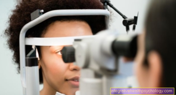
1. The organ of vision
The eye is responsible for conveying visual impressions from the environment to the brain. The eye is located in the eye socket formed by the skull of the face.
For more information, see: eye
Cornea
The approx. 600 micrometer thin Cornea (Cornea) covers the anterior segment of the eye. Together with the tear fluid, it forms the smooth, light-refracting surface of the organ of vision.
The cornea has one own Refractive power, with which it contributes to the mapping of visual stimuli on the retina.
She also has one protective Function by using the Intraocular pressure "cushions".
For more information, see: Cornea
iris
The iris (Iris) has roughly the same function as the aperture of a camera: it regulates the aperture by changing its size Incidence of light in the eye.
In its center it has an opening: this is it pupil.
By the amount of that stored in the iris Pigments (Dye) is the Eye color of man determined.
For more information, see: iris
pupil
The pupil represents the middle of the iris (rainbow skin): The ambient light reaches the inside of the eye through the pupil and creates the visual impression on the retina.
The pupil is enlarged or reduced in size via the muscles of the iris. This regulates how much light gets into the eye.
For more information, see: pupil
lens
The lens lies behind the pupil and, together with other structures, is responsible for refracting the incident light beam.
It is elastic and can be over Musculature actively arched.
In this way, the refractive power can be adapted to the various requirements.
For more information, see: Lens of the eye
Retina
The Retina consists of several layers and contains cells that receive light stimuli, convert them and pass them on to the brain via the optic nerve.
It is responsible for color and brightness vision:
The retina contains different cells for different colors and light intensities, which convert the light stimuli into electrochemical stimuli.
For more information, see: Retina and See
Blind spot
As blind spot is an area in the visual field of the eye where there are no sensory cells.
So it is a naturally occurring visual field loss.
Anatomically, the blind spot is created by the Optic nerve pupil described, i.e. the place where the Optic nerve leaves the eye.
For more information, see: Blind spot
Tear ducts
The Tear fluid serves to constantly moisten the eyes. Tears are very important for eye function.
The tears are from the Lacrimal gland produced, which sits on the upper outer edge of the eye.
From here the tears are spread over the whole eye by the blink of an eye.
At the inner corner of the eye the tears are then over the so-called Teardrop resumed and transported to the tear sac through the tear ducts. This empties into the nose.
For more information, see: Tear ducts
2. The organ of hearing
Outer ear
The outer ear is the first instance of the sound conduction apparatus and is used to receive and transmit the sound stimulus.
They belong to him auricle, the Ear canal and the eardrum.
For more information, see: Outer ear
- Ear canal
The human external auditory canal is approximately 2-2.5 cm in length.
It conducts the sound stimuli from the auricle to the eardrum.
In the first third of its course, its wall is formed by cartilage, the remaining two thirds are bony.
For more information, see: Ear canal
- eardrum
The eardrum is almost oval and is kept under tension by a cartilage ring.
It represents the boundary of the outer ear and middle ear.
Sound waves that hit the eardrum move it into vibration. This vibration is transmitted to the middle ear via the hammer handle, which is fused with the back of the eardrum.
For more information, see: eardrum
Middle ear
As Middle ear is the name given to the air-filled space that lies between the eardrum and the inner ear.
In it is the ossicular chain, consisting of a hammer (lat .: Malleus), Anvil (lat .: Incus) and stirrups (lat .: Stapes).
They are connected to one another in an articulated manner and mechanically transmit the vibration of the eardrum (i.e. the sound stimulus) to the inner ear.
For more information, see: Middle ear
Inner ear
The one lying inside the petrous bone Inner ear includes the auditory and equilibrium organs.
The Cochlea represents the hearing organ: it contains the various receptor cells (the so-called Organ of Corti), which make the sound stimulus perceptible for the brain.
The Balance organ lies above the cochlea and is organized in the form of several semicircular canals filled with fluid.
For more information, see: Inner ear
3. The olfactory system
nose
The nose consists of a bony and a cartilaginous part.
The bony part is called Root of the nose or nasal pyramid and represents a kind of foundation for the cartilaginous part of the nose that sits on it.
It is made up of the frontal bone, the maxillary bone and the Nasal bone educated.
The cartilaginous, mobile part of the nose consists of several different cartilages (triangular cartilage and nasal tip cartilage) that together enclose the nostrils.
The inner nose (also called the nasal cavity) connects to the outer nose.
For more information, see: nose
Nasal cavity
The nasal cavity is part of the upper airway and lies between the nostrils and the throat. It is further subdivided into the nasal vestibule and the main nasal cavity.
In addition to the respiratory function, it is relevant for html / antibiotika.htmlantibacterial defense, language formation and Olfactory function.
It stands in with different structures Skull area in connection.
For more information, see: Nasal cavity
Nasal septum
The Nasal septum divides the main nasal cavities into a left and a right side. The nasal septum thus forms the central border of the nostrils.
It forms with a posterior bony, a middle cartilaginous and an anterior membranous part with the nostrils externally visible shape of the nose.
For more information, see: Nasal septum
Sinuses
The sinuses are air-filled spaces that lie around the nose in the bones of the face.
They include:
- the Maxillary sinuses
- the Frontal sinus
- the Ethmoid sinus
- and the Sphenoid sinus
The paranasal sinuses serve to warm and humidify the air and act as a resonance space for improved voice and speech formation.
For more information, see: Sinuses
Nasal mucosa
The Nasal mucosa is a thin layer of tissue that is ours Nasal cavities lined from the inside.
It is made up of certain skin cells that are short Cilia is provided.
Additionally are in the mucous membrane Glands for secretion formation and venous plexus for Airflow regulation stored.
In addition, it contains the receptor cells that make the Smell enable.
For more information, see: Nasal mucosa
The nervous system
The Nervous system is a superordinate switching and communication system that is present in all more complex living beings.
The nervous system has the task of receiving information and forwarding it to the right places. It represents, so to speak, the cabling in our network.
It consists of several parts:
- the Neurons and Nerve fibers are the smallest units of the nervous system
- the Central nervous system (CNS), consisting of Spinal cord and Brain, is used for the integration and higher-level control of the neural information
- the Autonomic nervous system is not controlled arbitrarily: It works "autonomously" and regulates many processes in the human body
- the peripheral nervous system serves to forward and transmit stimuli from the body periphery to or from central interconnection points
For more information, see: Nervous system
Chapter overview
- Build up the nerves
- Central nervesystem
- Autonomic Nervous System
1. Structure of the nerves
Nerve cell
Neurons are nerve cells that specialize in stimulation generation and conduction.
As such, they form that smallest functional element of the nervous system.
A stimulus that hits a nerve cell causes an excitation that spreads in the cell membrane of the neuron and a so-called Action potential triggers. This is over long cell extensions that Axons, forwarded.
For more information, see Nerve cell
Motor neuron
As Motor neuron are specialized nerve cells that conduct nerve impulses to muscle fibers.
So you are responsible for coordinating and executing movements.
A distinction is made depending on the localization upper and lower motor neurons.
For more information, see Motor neuron
Axon
The term Axon refers to the tubular extension of a neuron through which the impulses formed in the cell are transmitted.
It takes its origin directly below the Nerve cell body (Soma).
The axon is either exposed or surrounded by a special layer of fat called the myelin sheath.
For more information, see Axon
Myelin sheath
The Myelin or Medullary sheath surrounds most of the nerve cells in the human body.
Similar to the sheathing of power cables, they serve the electrical insulation the nerve fiber. This allows impulses to be conducted faster and more safely.
From an anatomical point of view, they are formed by the cell membrane of certain cells that spiral around the axons.
For more information, see Myelin sheath
dendrit
Dendrites are, like axons, nerve processes of a nerve cell.
However, these do not move to the periphery, but serve to receive stimuli from upstream nerve cells.
They have a large number of branches.
For more information, see dendrit
Synaptic cleft
The synaptic cleft is the space between the end of a nerve cell and the corresponding target organ, such as other nerves or muscles.
Here the nerve impulse is modulated and transmitted in various ways.
For more information, see Synaptic cleft
Motorized end plate
The motorized end plate represents a special form of the synapse.
Here, released neurotransmitters (the Acetylcholine) Transmitting nerve impulses from a nerve cell to a muscle fiber, causing it to contract voluntarily.
For more information, see Motorized end plate
2. Central nervous system / CNS
The CNS (Zentrales Nervensystem) consists of the brain (cerebrum, encephalon) and the Spinal cord (medulla spinalis).
For more information, see: CNS / Central Nervous System
Cerebrum
The Cerebrum (lat .: Telencephalon) is the largest part of the human brain and is located just below the surface of the skull.
Its surface is heavily grooved, which gives it a characteristic appearance.
It is further divided into the bark (lat .: Cortex), in which the nerve cells of the brain are located, and that mark (lat .: Medulla), in which mainly the nerve tracts are located.
The cerebrum also includes some areas in which special interconnection processes take place:
- Basal ganglia
The term "Basal ganglia"refers to the core areas located under the cerebral cortex, which are primarily responsible for controlling motor processes.
For more information, see: Basal ganglia
- Limbic system
The Limbic system represents a functional unit of the brain that is used to process emotional impulses.
In addition, it controls the development of instinctual behavior and has a share in intellectual performance.
For more information, see: Limbic system
- Visual center
The Visual center is lying in Occipital lobe (Posterior lobe) of the cerebrum. This is where all the information gathered through the eye arrives, is processed and made "conscious".
For more information, see: Visual center
Meninges
The brain is controlled by the so-called Meninges surround. This consists of several layers, of which the outer one lies directly on the skull bone.
The meninges protect and supply the brain.
For more information, see: Meninges
Diencephalon
The Diencephalon is a part of the brain that is located between the cerebrum and the brain stem.
It consists of:
- Thalamus
- Epithalamus (epi = on it)
- Subthalamus (sub = under) with Globus pallidus (Pallidum)
- Hypothalamus (hypo = below)
In the diencephalon are mainly Stimuli of the sense organs processed and forwarded accordingly.
For more information, see: Diencephalon
Brain stem
The Brain stem of the brain includes that Midbrain, the bridge, the Cerebellum as well as the extended markwhich in the Spinal cord transforms.
In addition, the brainstem contains the nuclei of the third through the twelfth Cranial nerves.
Overall, that is Brain stem responsible for the regulation of vital processes such as sleep, breathing, Blood pressure level and urination.
For more information, see: Brain stem
- Cerebellum
The Cerebellum (lat .: Cerebellum) lies in the posterior fossa under the cerebrum.
It can be roughly divided into 2 hemispheres, which are separated by what is known as the worm, an elongated section of the cerebellum.
The surface The cerebellum is enlarged by innumerable folds to provide more space for nerve cells and fibers.
The Function of the cerebellum includes, in a nutshell, the control of motion sequences.
For more information, see: Cerebellum
- Extended mark
The extended mark (lat .: Medulla oblongata) is the furthest down (caudal) located part of the brain.
It contains Nerve nuclei and -tracksthat control vital processes such as breathing.
The Medulla Reflex centers for Reflexes such as sneezing, coughing, swallowing, and vomiting.
For more information, see: Extended mark