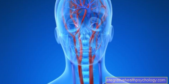
Carotid artery
General
Three different arteries are commonly referred to as the carotid artery. On the one hand the big common one Common carotid artery and the two arteries arising from it, Internal carotid artery and External carotid artery.

Common carotid artery
The Common carotid artery, also called "carotid" resp. Carotid artery is the common carotid artery.
Since they are in the Depth of the neck runs and in its course Food- and windpipe accompanied by the chest towards the head, it is also known as the carotid artery. your Pulse is easily palpable on the neck.
She runs paired on both sides of the neck and arises on the right side from the Truncus brachiocephalicus, on the left mostly directly from the Aortic arch.
In humans it divides into the "Carotid fork" into an outer and an inner artery. The height of the carotid fork is individually different and can between the second and sixth cervical vertebrae lie. In most people, it is at the level of the fourth cervical vertebra.
At the exit of the inner carotid (Internal carotid arterya) lies the Sinus caroticus. This one is with Pressure receptors (Baroreceptors) equipped and monitors the Blood pressure in the arterial system. From here the information about the pressure is sent to the brain and the heart forwarded. In addition, certain measure in this area Chemoreceptors the level of carbon dioxide (CO2), oxygen and the pH value in the blood.
Internal carotid artery
The Internal carotid artery, also called the internal carotid artery, is one of the vessels supplying the brain of the human. In addition, it supplies via the Ophthalmic artery in humans that eye With oxygenated blood.
The course of the internal carotid artery is divided into four parts. The neck part (Pars cervicalis) ranges from their departure from the large Common carotid artery until they join the Skull base.
At the beginning it is usually behind the smaller external carotid artery (External carotid artery) and then continues towards the center where it reaches the base of the skull.
In this neck part there is the Internal carotid artery no branches. The closes at the neck part Petrous part (Pars petrosa) at. It runs there in the temporal bone and first pulls further upwards before being in the anterior wall of the Tympanic cavity draws an arc and then direction Sphenoid body runs. This bow is also called Carotid knee designated.
The Pars petrosa gives different branches to the tympanic cavity (Caroticotympanic arteries) and Pterygoid Canal (Arteria canalis pterygoidea) from. In the area of the inner opening of the carotid canal, the internal carotid artery is often only separated from it hard meninges (Dura mater) covered.
Right on the Inside of the base of the skull the carotid runs through the Cavernous sinuswhich is why this part is called Pars cavernosus referred to as.
In this area, the artery makes another S-shaped curve from the back bottom to the front top. This is called Carotid siphon designated. In this part the carotid gives off branches Neurohypophysis (Inferior hypophysial artery), to the Trigeminal ganglion (Rami ganglionares trigeminales), to the meninges (Rami meningeus) and Cavernous sinus (Rami sinus cavernosi) from.
After breaking through the hard meninges, the carotid goes into theirs "Brain part“ (Pars cerebralis) above. This part is in the Subarachnoid space at the base of the brain. In this section it runs from the back bottom to the top front and immediately afterwards releases its branch to the eye (Ophthalmic artery).
Most of the time this part also goes out Posterior communicating artery out which part of the Circulus arteriosus cerebri and connects the anterior and posterior flow areas in the brain.
After submitting the Anterior choroid artery, which supplies various brain structures, the internal carotid artery divides into the anterior (Anterior cerebral artery) and the middle (Media cerebral artery) Cerebral artery. These two arteries supply a large part of the Cerebrum.
Segments of the internal carotid artery
The internal carotid artery can be divided into 4 sections subdivide:
Pars cervicalis (Neck part): It starts at the carotid sinus and runs through the carotid canal into the base of the skull.
Pars petrosa (Temporal bone): It pulls up through the temporal bone into the tympanic cavity, where it makes an arc forward, also known as the carotid knee. It is in close proximity to the venous plexus.
Pars cavernosa: It runs along the inside of the skull base and through the cavernous sinus.
Pars cerebralis: It runs in the subarachnoid space at the base of the brain from back to front.
There is also a second classification based on clinical criteria. The cerebral and cavernous pars are also divided into segments C1-5.
The external carotid artery cannot be divided into sections.
Branches of the internal carotid artery
The internal carotid artery has 4 sections:
- The Pars cervicalis does not give off branches.
- The Pars petrosa gives off the ramus caroticotympanicus (tympanic cavity) and the artery canalis pterygoidei (canal).
- The Pars cavernosa divides into 6 branches: R. tentorii basalis, R. tentorii marginalis, R. meningeus (meninges), R. sinus cavernosi (sinus), A. hypophysialis inferior (pituitary gland) and R. ganglionis trigeminalis (ganglion trigeminale).
- The Pars cerebralis there also 7 branches from. The R. clivi, the A. hypophysialis superior (pituitary gland), the A. ophthalmica (eye) and the A. choroidea anterior are classic arteries. The posterior communicating artery, the media cerebral artery and the anterior cerebral artery, on the other hand, form parts of the Circulus arteriosus. This is a circular anastomosis that divides the flow areas of the Aa. carotis and the aa. vertebralis connects and should create a certain balance in case of insufficient blood flow.
Supply areas of the internal carotid artery
The internal carotid artery supplies large parts of the Brain (A. cerebri media and anterior, A. hypophysialis, A. choroidea anterior, ...). Especially the front part and gives branches to eye (A. ophthalmica), to the trigeminal ganglion, to the tympanic cavity, to the nose and to share the forehead from.
Together with the vertebral artery it forms the Circulus arteriosus.
External carotid artery
The external carotid artery supplies the soft tissues and bones of the skull, as well throat, Larynx, thyroid and the hard meninges.
She goes to the Carotid fork from the Communicating carotid artery and is usually the smaller artery of the two Carotid arteries.
It usually lies in front of the internal carotid artery and runs between the Stylopharyngeal muscle, the Stylohyoid muscle and the rear part of the Digastric muscle.
In its course it will be dated Hypoglossal nerve over and from Glossopharyngeal nerve undercrossed. in the Jaw angle it divides into its terminal branches: Maxillary artery and Superficial temporal artery.
Numerous branches emerge from these two branches to the various structures of the skull and neck emerged. This is how the External carotid artery about the Lingual artery and the Ascending pharyngeal artery the tongue and the throat.
About the Superior thyroid artery will the thyroid With Blut supplied and about the Facial artery and its terminal branches the face. The Occipital artery takes care of the back of the head that Posterior auricular artery the back of the Ear and the Maxillary artery the jaw. Numerous small branches emerge from these larger branches, which supply the soft tissues of the skull and neck.
Branches of the external carotid artery
The external carotid artery gives off 3 anterior branches: the A. thyroidea superior (Thyroid, larynx), the A. lingualis (Floor of the mouth, tongue) and the Facial artery (superficial face).
Also there is a medial branch that A. pharyngea ascendens (pharynx to skull base), ab and two posterior branches, the Occipital artery (Occiput) and the A. auricularis posterior (Ear region).
Finally she gives with the A. maxillary (Masticatory muscles, posterior inner part of the facial skull, meninges) and the Superficial temporal artery (Temporal region, part of the ear) two terminal branches.
Supply areas of the external carotid artery
The external carotid artery supplies the outer skull and parts of the neck. It gives off branches to the larynx, thyroid gland (superior thyroid artery) and pharynx (ascending pharyngeal artery). In the mouth area, it supplies the Floor of the mouth, the tongue (A. lingualis) and the Masticatory muscles (Maxillary artery).
In addition, it gives off branches to the superficial face (A. facialis), to the temporal region (A.temporalis superficialis), to the occiput (A. occipitalis) and also to part of the ear. It also supplies parts of the inside of the skull, such as the base of the skull, the rear inner part of the facial skull and the meninges.
Aneurysm
An aneurysm is one Weak point in the wall of a vessel. Usually an asymptomatic bulge occurs first, which is often discovered by chance. However, if the vessel ruptures due to a sudden increase in blood pressure, it can close life threatening bleeding lead in the skull.
The aneurysms often occur on the arteries of the circulus arteriosus. They are mostly located on the anterior communicating artery, but the media cerebral artery, the internal carotid artery and the posterior communicating artery are also often affected.
If the aneurysm ruptures, it will bleed into the space between the arachnoid and the inner meninges. Typical symptoms are very strong, suddenly appearing, so-called Annihilation headache and Neck stiffness.
Bleeding in the skull must be recognized and treated as quickly as possible, otherwise the increase in volume caused by the bleeding will put pressure on the brain and shift the brain tissue to the opposite side. As a result, important structures can be squeezed or kinked and infarcts (tissue collapse) can occur in the affected areas. Depending on the area, this has considerable effects (e.g. motor failures) and can often lead to death. Therefore, early detection and therapy before the rupture are essential for a good prognosis.
Narrowing of the carotid artery
A narrowing or obstruction of part of the internal carotid artery can usually occur for two reasons. Either has a Blood clots replaced and has become a embolism (Vascular occlusion) or the vessel is atherosclerotically changed and it's gotten so over time at this point thrombus educated.
Most blood clots that block blood vessels in the brain come either from the left atrium of the heart (for example, due to valve disease) or from the carotid bifurcation. These clots enter the brain with the bloodstream and settle in the places where they can no longer proceed due to the small diameter of the vessels. The media cerebral artery is most commonly affected.
The closure of a vessel leads to circulatory disorders and thus to a lack of oxygen and nutrients in the area that is no longer supplied. This ultimately leads to the so-called stroke. Those affected often have various types of neurological deficits. Typical symptoms are paralysis, speech disorders, visual field defects or numbness in parts of the body.


.jpg)