CT abdomen
What is an abdomen CT?
CT is the so-called computed tomography. This is a procedure that works just like the classic X-ray examination with X-rays. However, not just one image is recorded, but a series of images while the computer tomograph rotates around the patient.
With a CT abdomen, only the patient's abdomen and pelvis are examined. Such an examination can be necessary for various illnesses and injuries in the abdominal area and is completely painless. It also provides an extremely accurate representation of the internal organs and structures.

Preparing an abdomen CT
Unless it is an emergency, a CT abdomen examination is planned and the patient is invited to a preparatory meeting.
Some important points should be clarified. Many patients already have known pre-existing conditions, such as high blood pressure or diabetes mellitus.
These must be known to the doctor before the examination. Thyroid or kidney diseases and allergies are particularly important in the context of the CT examination, as these can become problematic when contrast media are administered.
An up-to-date list of medications should also be made available to the doctor. In addition, there is a possible pregnancy in women, as this excludes a CT examination in most cases.
If the examination requires contrast medium which the patient must drink, the doctor will inform the patient when and at what intervals this should be done.
Read more on the subject at: Computed Tomography
Do you have to be sober to get an abdomen CT?
Whether and how much can be eaten before the examination depends entirely on the region to be examined. If the gastrointestinal tract is to be examined, it is often necessary to avoid food for 8 hours before the examination. When assessing the urinary tract and the bladder, however, a light meal may be consumed before the examination.
In general, alcohol should be avoided on the day of the examination. Some medications may also not be taken before the examination because of a possible administration of contrast medium. However, this will be discussed in a preliminary discussion with the attending physician.
The procedure of the CT abdomen
The CT abdomen itself is very quick and completely painless. The patient lies down on a special couch that can be moved into the computer tomograph. If the examination requires contrast agent, which is intended to represent the vessels, this is administered to the patient in the vein shortly before the examination begins.
The X-ray assistants leave the room for the actual examination. You can give instructions to the patient over an intercom. For the examination of the abdominal cavity, the patient lies flat on his back in most cases and is not allowed to move. The couch is moved into the device and circled by it.
For some recordings it is necessary to breathe in and / or hold your breath for a certain period of time so that the organ to be examined is represented well and the images do not blur. When and for how long this should happen, the patient is always informed during the examination. At the end of the examination, the X-ray assistants re-enter the room. The CT machine no longer emits any X-rays.
The evaluation of the CT abdomen
The evaluation of the CT images is carried out by a radiologist who has previously been informed of the reason for the examination by the attending physician (e.g. abdominal pain of unknown origin). He then assesses the recordings made with regard to the patient's symptoms. A possible cause can often be found quickly, e.g. if there are stones in the gallbladder or kidney.
The evaluation is then discussed again with the attending physician and the further procedure and possible therapies are clarified with the patient.
What can you see in the evaluation?
The radiologist who evaluates the CT images usually receives them in digital format. The images can then be looked through continuously on the screen, as if working your way through your body from top to bottom. The extent and location of the individual internal organs can be assessed. For example, a malformation of organs can be shown very well.
In addition, the structure and density of the organs can be determined using the various gray levels. This is very important for tumors, for example. These often have a different structure than the surrounding tissue and are noticeable through different shades of gray. The same applies to stones, which are often found in the urinary tract. These often appear as clearly visible white spots. Air or liquid in the wrong places in the body is also noticed early.
Computed tomography risks
The CT in general creates a higher radiation exposure than a conventional X-ray examination. Radiation-sensitive organs such as the stomach or ovaries in women are particularly located in the abdomen and pelvic area.
It is therefore important to ensure that the CT examination, especially the CT abdomen examination, is only carried out when it is sensible and necessary.
CT examinations should be used sparingly, especially in children, for whom the radiation is more dangerous than for the elderly. Nevertheless, the risks of CT should not be placed above the benefits.
This is because such an examination is absolutely helpful in the emergency area and can save the patient's life by quickly identifying potentially life-threatening conditions.
How high is the radiation exposure?
The radiation exposure of humans is usually given in Sievert or Milli-Sievert (mSv). In order to have a comparison value, the radiation that humans are naturally exposed to during the year, e.g. by cosmic radiation, given with approx. 2-2.4mSv.
The CT abdomen examination is one of the more radiation-intensive examinations. On average, a person is exposed to about 10 to a maximum of 20mSv.
However, this value cannot be compared completely with the above, since the natural radiation affects the entire body, while the CT examination only exposes a small part of the body to additional radiation.
For details, see Radiation exposure in computed tomography.
The duration of the investigation
In contrast to an MRI examination, a CT examination is very quick.
The examination itself usually only takes a few minutes. However, it must be taken into account that contrast media is required for many examinations. In the abdominal area in particular, the contrast agent has to be drunk frequently and then, depending on which organ is to be assessed, it takes one to two hours to get to the destination.
It should also be noted with the CT abdomen that several series of images may have to be taken. Overall, however, the examination rarely takes more than 20-30 minutes.
How high are the costs?
The examination using CT is one of the more expensive examinations. According to the GOÄ (Fee Regulations for Doctors), the simple rate for a CT examination in the abdominal area is € 151.55.
This is calculated for patients with statutory health insurance. For privately insured patients, 1.8 times the rate can be charged. In the present case, this amounts to € 272.79.
Do i need contrast media?
The question of whether a contrast agent is necessary for the CT abdomen examination also depends on the type of examination. However, this is very often the case in the abdomen or pelvis. Because both the digestive organs and the urinary tract are shown much more precisely when contrasting is carried out. In the area of the gastrointestinal tract, this can sometimes even be sufficient just by drinking water.
Then, shortly before the examination, about half a liter of water must be drunk in a short time. Contrast agent in the vein (IV contrast agent) is mainly used when blood vessels are to be assessed, but the excretion of the contrast agent via the urinary tract can also be precisely assessed.
Read more on the topic: Contrast media
What is a LowDose CT
A low-dose CT is a CT examination that has a lower radiation risk than a conventional examination.
This is particularly often used in the search for urinary stones in the context of colic. Precisely because urinary stones often occur repeatedly, it is important to keep the radiation as low as possible through the CT examinations performed. However, the type of CT examination carried out to search for stones is usually very radiation-intensive.
With the help of the LowDose technology, the radiation can be reduced to that of a conventional overview image of the abdominal cavity. In this way, the risk of the patient suffering from radiation damage later on can be minimized.
Read more on the subject at: Low-dose CT
Which organs do you see?
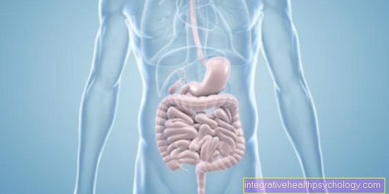
The organs of the digestive system can be assessed during a CT abdomen examination. These include the esophagus, stomach, small intestine, and large intestine. The spleen, pancreas, liver and gall bladder can also be visualized with the CT scan.
In the area of the gallbladder, gallstones in particular can be seen very clearly.
Another important area is the urinary system. In addition to the kidneys and ureters, the bladder can also be seen here.
In addition to the organs, vessels, especially the arteries, can also be assessed. Especially the large vessels in the abdomen, e.g. the aorta, can be examined for bulges (aneurysms).
What is a Hydro CT abdomen?
A hydro-CT describes a special type of examination in which water is used as a contrast medium. The stomach and intestines in particular can be assessed well with a hydro-CT. To do this, the patient must drink approx. 500 ml of water shortly before the start of the examination. In addition, an anticonvulsant drug is often administered, which lowers gastrointestinal activity.
The water acts as a so-called negative contrast medium, i.e. it stains the stomach and intestines darkly in the images. As a result, the wall of the organs can be shown particularly well and abnormalities can be better recognized.
Do I have to take off my clothes for the examination?
Normally, only the clothes that are in the examination area have to be removed. This is particularly necessary because of metallic buttons and zippers, as these can interfere with the recordings. During an abdominal CT, the trousers usually have to be taken off, and women sometimes have to take off their bra.
In some clinics or practices, however, trousers or shirts can also be left on. However, this must be clarified individually with the respective practice or clinic.


