The inner ear
Synonyms
Latin: Auris interna
English: internal ear
definition
The inner ear is located inside the petrous bone and contains the hearing and balance organs. It consists of a membranous or membranous labyrinth, which is surrounded by a similarly shaped bony labyrinth.
Anatomy and function

The hearing organ:
The cochlea is part of the hearing organ / ear in the inner ear (Cochlea).
It consists of the cochlear Labyrinth with a membranous spiral duct (Chochlear duct). It contains the sensory epithelium with two different receptor cells, the so-called Corti-Organ. The tip of the snail points to the side forwards and not upwards.
The bony snail duct (Canalis spiralis cochleae) in the inner ear is about 30-35 mm long. It wraps around the approximately 2.5 times Modiolus, its bony axis, which is penetrated by several cavities and that Ganglion spiral (Nerves for the impulse reception of the frequencies) contains. The basal cochlea of the inner ear is from the tympanic cavity (middle ear) as a protrusion (Promontory) to recognize.
The membranous compartments are storied in cross section. Above and below are with Perilymph (Ultrafiltrate of the blood plasma; resembles the extracellular fluid) filled spaces: the Scala vestibuli and the Scala tympani. In the middle of the inner ear there is another space, the Cochlear ductwhich with Endolymph (resembles the composition of the intracellular fluid) is filled. It ends blindly towards the tip of the snail, while the Scala vestibuli and Scala tympani at the snail hole (Helicotrema) are connected to each other at the tip of the snail in the inner ear. In the cross-section the Cochlear duct triangular and is separated by the so-called Reissner membrane Scala vestibuli and through the basilar membrane from the Scala tympani Cut. On the side wall there is a particularly metabolically active area (Stria vascularis) who the Endolymph secreted.
The Basilar membrane arises from a bony protrusion and becomes wider and wider from the base of the snail to the tip of the snail. This is where the sensory apparatus is found with the inner and outer hair cells, which have a ratio of 1: 3. The hair cells wear different lengths Stereovilli. The smallest of them are connected to one another by protein threads. This is where an external stimulus is converted into a physiological signal (Transduction) take place via certain ion channels. The Corti-Organ is used by the Tectorial membrane covered. At rest, i.e. without external stimulus, only the outer hair cells in the inner ear touch the tectorial membrane. Fibers of the auditory nerve close to the inner hair cells (Cochlear nerve), which forwards the information to the brain. The function of the hearing organ is to convert the incoming sound waves into electrical impulses. The exact transduction processes and the principle of sound conduction are described below.
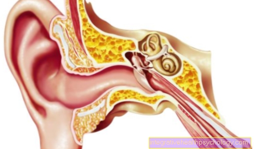
Figure ear

A - outer ear - Auris externa
B - middle ear - Auris media
C - inner ear - Auris interna
- Ear strip - Helix
- Counter bar - Antihelix
- Auricle - Auricula
- Ear corner - Tragus
- Earlobe -
Lobulus auriculae - External ear canal -
Meatus acousticus externus - Temporal bone - Temporal bone
- Eardrum -
Tympanic membrane - Stirrups - Stapes
- Eustachian tube (tube) -
Tuba auditiva - Slug - Cochlea
- Auditory nerve - Cochlear nerve
- Equilibrium nerve -
Vestibular nerve - Inner ear canal -
Meatus acousticus internus - Enlargement (ampoule)
of the posterior semicircular canal -
Ampulla membranacea posterior - Archway -
Semicircural duct - Anvil - Incus
- Hammer - Malleus
- Tympanic cavity -
Cavitas tympani
You can find an overview of all Dr-Gumpert images at: medical illustrations
Transduction processes and the principle of sound processing in the inner ear
The im Inner ear incoming sound is transmitted via the outer ear to the eardrum directed. There the resulting vibrations are transferred to the ossicular chain hammer, anvil and stirrup in the Middle ear brought up to the oval window to the inner ear. The oval window adjoins the Scala vestibuli. The stapes footplate sets the inner ear fluid and the membranes of the cochlea in motion through continuous inward and outward movements. This is where the signal transduction process begins, which can be divided into 3 stages:
- Creation of a traveling wave
- Excitation of the outer hair cells
- Excitation of the inner hair cells by amplifying the traveling wave through the outer hair cells
1. Creation of a traveling wave:
A Traveling wave arises in the inner ear through undulating movements. It starts at the oval window and then runs the Scala vestibuli up to the snail tip. Would be cochlear Partition a homogeneous structure, a synchronous oscillation would occur. But their stiffness decreases from the screw base to the screw tip. It follows that the partition oscillates in the form of a traveling wave. Overall, there is an amplitude (oscillation) maximum for each frequency. If the excitation frequency of the external sound stimulus is equal to the natural frequency of the basilar membrane, an amplitude maximum follows. This principle of Frequency dispersion (Frequency-location mapping, spatial theory) allows a characteristic assignment of frequencies (Tonotopy). High frequencies are found at the base of the snail in the inner ear, while low frequencies are found at the tip of the snail in the inner ear.
2. Excitation of the outer hair cells
At the maximum of the wave movement, the Stereovilli of the outer hair cells bent the most. There is shear movement between the basilar and tectorial membrane. The tip-links are stretched or relaxed by upward and downward movements. This opens or closes ion channels in the inner ear and changes the potential of the hair cells. They then actively change their length and strengthen the traveling wave. The frequency selectivity is thus improved.
3. Stimulation of the inner hair cells
The inner hair cells in the inner ear are only excited by the amplification mechanism of the outer hair cells. Now they too come partially into contact with the tectorial membrane and the shear off of the Stereovilli ensures the release of a neurotransmitter at the base of the hair cell, which then the annoy of the auditory nerve (Cochlear nerve) excited. From here the information continues to the brain managed and processed.
The vibrations in the inner ear lead to the radiation of sound energy to the outside. The traveling wave continues from the Scala vestibuli over the snail tip to the Scala tympani, which ends at the round window. Sound coming from the ear can be measured as so-called evoked otoacoustic emissions. Emissions in the inner ear triggered by "clicks" can be recorded with a microphone and used for hearing screening, especially in newborns.
Summary
The Inner ear represents a complex structure with the help of which we can orientate ourselves in space. Sound perception also plays an extremely important role in our social coexistence.