Diagnosis of gallstones
How is the diagnosis of gallstones made

The doctor will first try based on a specific question (History taking) the causes of those described by the patient Pain to find out.
He'll likely ask the following questions:
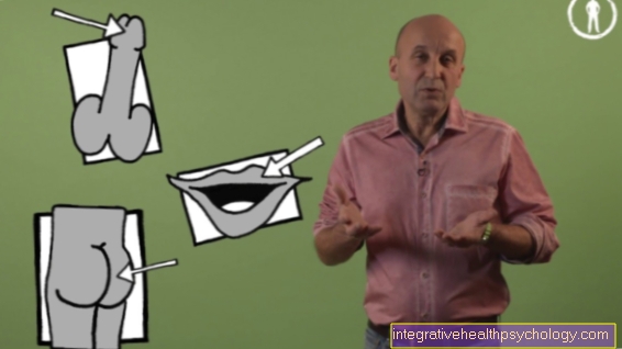
- When do the complaints occur?
- Does the pain occur mainly after and mainly after eating high-fat food?
- Does the pain occur at night while lying down?
- Is there nausea and vomiting?
- Is the pain radiating?
- Where is the pain localized?
- Is the pain undulating?
The doctor will now perform a clinical diagnosis of the patient's abdomen. In addition to checking the tenderness to pressure, the so-called Murphy characters to mention: after the patient exhales, the doctor deeply presses the area on the abdomen where the gallbladder is located and lets the patient inhale again. If he stops the inhalation maneuver and complains of pain, that speaks for you Gallstone in the gallbladder (Murphy's sign positive).
Next, the doctor gets one Ultrasound examination of the abdomen. It is an accurate and safe method of stone diagnosis. The one or the other Gallstones appear as a white structure with a subsequent acoustic shadow. In the ultrasound examination, the doctor can also determine whether the Gallbladder or the aisles are thickened. This would be for a Inflammation of the gallbladder (Cholecystitis) or biliary inflammation (Cholangitis) speak. A so-called high-frequency signal analysis can be used to assess whether the stone is a cholesterol stone and whether it is calcified. Can also be a Computed Tomography can be made to explain the composition of the stone.
In order to rule out that the symptoms are an inflammatory process when diagnosing gallstones, a blood test should be carried out on the patient. The white blood cells (Leukocytes) and the inflammatory proteins (CRP) whose increase would be typical of an inflammatory process. But also liver-, and bile values should be explored (gamma GT, alkaline phosphatase).
Another method of diagnosis, especially in the case of drainage problems caused by gallstones, is endoscopic retrograde cholangio-pancreatography (ERCP). Here, a hose with a camera at the tip is inserted into the stomach and then advanced into the duodenum. From there you can get into the bile duct and examine whether there is a gallstone in it.
A contrast medium is injected through the advanced instrument into the bile duct and then finished X-ray image gallstones can be seen, which obstruct the flow of the contrast medium. An even more precise representation of the gall bladder and the bile duct system can also be produced by an ultrasound probe located at the beginning of the tube.