The respiratory muscles
synonym
Auxiliary respiratory muscles
introduction
Respiratory muscles (or auxiliary respiratory muscles) are different muscles from the group of skeletal muscles that help expand or contract the chest. In this way, these muscles make an important contribution to inhalation and exhalation.
By far the most important component of the respiratory muscles is the diaphragm (lat. Diaphragm). In addition, various muscles of the chest, abdomen and back are included in the group of respiratory muscles. A broad distinction is made between so-called chest breathing and abdominal breathing.

Inspiratory respiratory muscles
The most important component of the respiratory muscles during inspiration (inhalation) is the so-called diaphragm (Diaphragm). For this reason, diseases of the diaphragm can also cause breathing-dependent pain. Basically, this structure is less of an actual muscle than a plate made up of muscle fibers and tendons.
The diaphragm is about 3 to 5 mm thick in humans and separates the thoracic from the abdominal cavity.
The diaphragm alone, as a respiratory muscle, can provide between 60 and 80 percent of the muscle work required for inhalation through sufficient contraction. For this reason, the diaphragm is considered to be the "motor" of so-called diaphragmatic or abdominal breathing.
In a neutral breathing position (i.e. at the end of expiration), the diaphragm assumes a position bulging towards the chest. When you begin to inhale, the muscle-tendon plate begins to shorten by up to 35 percent. In the course of this, there is a significant flattening and the formation of a cone shape. Through this process and the interaction with other components of the respiratory muscles, the chest cavity is greatly enlarged.
At the same time, the activity of the diaphragm triggers an increase in the negative pressure within the pleural space.
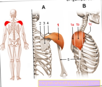
In addition to the diaphragm, the external intercostal muscles (External intercostal muscles), the scalene muscles and the muscles in the area of the costal cartilage (Intercartilaginous muscles) belongs to the group of inspiratory respiratory muscles.
More information can be found here:
- The chest breathing
- Diseases of the diaphragm
Auxiliary respiratory muscles during inhalation
The diaphragm is the most important respiratory muscle, but with more intense stress, the oxygen demand can often only be met with the aid of the auxiliary respiratory muscles. This particular group of respiratory muscles is mainly attached to the bony structures of the chest. For this reason, the individual muscles enable an enlargement of the chest cavity and a significant increase in breathing volume.
In contrast to the normal respiratory muscles, the auxiliary respiratory muscles can neither be used for inspiration nor expiration support in the normal body position. The activation of these special respiratory muscles requires the reversal of the insertion and origin of the muscle fibers. In order to be able to use the auxiliary respiratory muscles for support, it is usually sufficient to press the arms firmly against the thighs, a table or the like.
This group of respiratory muscles therefore plays a decisive role in sports activities. In addition, it makes breathing easier in the presence of various lung diseases. The diseases relevant in this context include water retention in the lungs (pulmonary edema), asthma and scarring of the lung tissue (pulmonary fibrosis).
The most important representatives of this group include the respiratory muscles
- the rib muscles (Levatores costarum muscles),
- the front saw muscle (Serratus anterior muscle),
- the posterior upper and posterior lower saw muscle (Serratus posterior superior and inferior muscle),
- the pectoralis major and minor (Pectoralis minor et major muscle) and
- the muscle between the sternum and mastoid (Sternocleidomastoid muscle).
Expiratory respiratory muscles
During intense physical activity and / or the presence of various lung diseases, the so-called expiratory muscles can be used to intensify the normally passive exhalation process.
The most important breathing muscles in exhalation are
- the internal intercostal muscles (Inercostales interni et intimi muscles),
- the subcostal muscle (Subcostal muscle)
- and the transverse lower thoracic muscle (Transversus thoracis muscle).
The activation of this part of the respiratory muscles is usually controlled by an increased consumption and need for oxygen at the brain level. Depending on the load situation, a different number of these muscles can be used with different intensities.
Auxiliary respiratory muscles during exhalation
Breathing support at the expiration level is primarily provided by muscle groups in the abdomen and back.
The most important auxiliary expiratory muscles are
- the transverse external abdominal muscle (External obliquus abdominis muscle),
- the transverse inner abdominal muscle (Internus abdominis oblique muscle)
- and the horizontal lower abdominal muscle (Transversus abdominis muscle), which mainly contributes to the intensification of exhalation during exertion and / or lung diseases.
If, despite the increased activity of the respiratory muscles, the increased oxygen consumption cannot be covered, the large back muscle (Latissimus dorsi muscle) have a supportive effect. This muscle is also able to make it easier to cough up stuck secretions. It is therefore not just a part of the respiratory muscles, but also a so-called cough muscle.
The lumbar square muscle (Quadratus lumborum muscle) belongs to the group of the expiratory auxiliary respiratory muscles.
Pain in the respiratory muscles
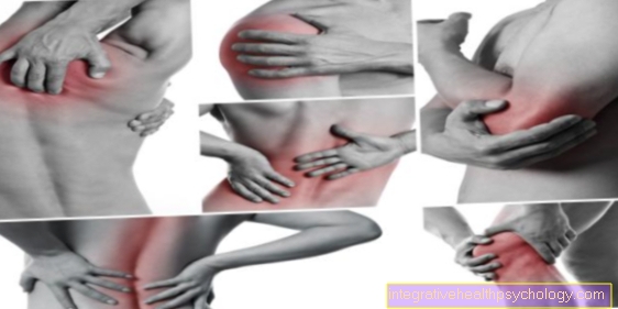
Depending on the location and on the inspiration or expiration, pain in the respiratory muscles can have a variety of causes.
- If there is pain in the chest when inhaling, tension in the intercostal muscles may be a reason.
- If the painful area is more concentrated at one point on the costal arch, a rib bruise or rib fracture could be the reason.
- If the symptoms are more towards the abdomen, it can be harmless air-filled loops of the intestine, tension in the abdominal muscles or serious causes such as swelling of the liver or spleen.
- A defect in the diaphragm, such as a hernia, can also cause pain.
If the pain persists longer, you should consult a doctor to clarify the symptoms.
Please also read our articles on this:
- Bruised rib pain - what can I do?
- Symptoms of a broken rib
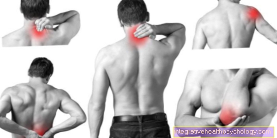
Symptoms of tight breathing muscles
Tense breathing muscles can make themselves felt through various symptoms.
- If you feel pain while breathing, you automatically breathe in and out less deeply. One becomes more short of breath and subjectively perceived shortness of breath can arise. This could be remedied by deep breathing, but since it hurts to breathe, a vicious circle begins.
- If necessary, one takes a relieving posture, such as a curvature of the upper body. Tense neck, shoulder or abdominal muscles can also be signs of tense respiratory muscles.
- If you have got used to an unconscious bad posture while breathing, such as breathing with your stomach pulled in and shoulders pulled up, the neck muscles and abdominal muscles can harden.
- If it hurts when breathing in the chest or if there is a feeling of tightness when breathing in, tense rib muscles can be behind it.
How do you loosen tight respiratory muscles?
Tense breathing muscles can be extremely painful. In order to release the tension, the muscles have to be stretched, which in turn causes pain, but is essential in order to achieve the pain-free initial state.
Even if it is uncomfortable at first, you should consciously relax during all exercises.
Various exercises help in stretching the respiratory muscles.
- One exercise is the pasha seat, in which you sit with your legs apart on a chair or armchair, lean against it and let your arms hang loosely. Then take a deep breath so that your chest rises and your stomach bulges, and then you relax. This exercise is repeated a few times so that the shoulder, neck and abdominal muscles can relax.
- Another possibility is to sit on a stool with your legs apart, bend your upper body forward, support your elbows on your thighs, let your head, arms and shoulders hang down and breathe in and out as deeply as possible. This exercise can also be performed while standing against a wall.
- If the muscles between the ribs are tense, you can try to stretch them by "sniffing" them. Like a dog, you "sniff" through your nose piece by piece and repeat the exercise a few times.
- If the hardening of the respiratory muscles is stubborn, you can also use fango packs and massages for relaxation.
What is respiratory muscle training and how is it done?
Respiratory muscle training or respiratory gymnastics serves to strengthen the respiratory muscles. Both the strength of inhalation and exhalation and endurance can be trained using various devices or exercises.
Not only people with shortness of breath or lung diseases use breathing gymnastics, healthy people also use this technique - for example singers and musicians who play a wind instrument.
Various devices aim to promote the strength of the respiratory muscles through resistance.
For example, a respiratory training device creates a pressure that initially makes inhalation impossible. If the patient breathes in harder on the device, the pressure is increased and above a certain threshold value a valve opens so that fresh air flows in. This training device is called "Threshold IMT" and is often used by COPD patients for exercise.
Another device lets the patient breathe out against resistance, so that specifically the muscles that are responsible for expiration are trained.
Read more about this: Breathing exercises