Tear ducts
Synonyms in a broader sense
Dacryocystitis, canaliculitis
introduction
The largest part of the tear system is located in the inner corner of the eye. It is made up of a tear-producing and a tear-transporting part.
The constant moistening of the surface of the eye is of great importance for the vision and function of our eyes.
The cornea, which is dependent on the supply of the nutrients contained in the tear film and the oxygen present in the air, benefits in particular. In addition to external nutrition, the cornea also has internal nutrition from the aqueous humor contained in the anterior segment of the eye.
In addition to nourishing and moisturizing the eye, the cleansing function is also very important. Everyone knows the phenomenon that the eyes begin to water heavily when a foreign body (e.g. dust particles) gets into the eyes.
Function of the tear ducts
The tear ducts serve to moisten the eye and its structures. To maintain and nourish the cornea, moistening by the tear film is of great importance. The components of the tear fluid supply the cornea with the necessary nutrients.
Since the cornea does not have its own nourishment, such as blood vessels, the tear film is essential. So that the eye does not dry out, it is continuously moistened by the tears. The distribution takes place via the blink of an eye.
Anyone who has ever suffered from "dry eyes" knows that too little tear fluid can be very uncomfortable. There are tear substitutes that can be used as eye drops for therapy.
You can find more on this topic under: Dry eyes
In addition, the flow of tears has a cleansing function by rinsing the eyes. If a foreign body (for example dust particles) gets into the eyes, the eyes quickly begin to water heavily.
This is used to rinse the eye and remove the foreign body. Incidentally, this foreign body always feels bigger than it really is. Even the smallest speck of dust causes severe pain and a foreign body sensation in the eye. The conjunctival sac, into which the tear fluid drains, is also cleaned in this way.
Tears also contain enzymes that have a bactericidal effect, i.e. can kill bacteria.
construction
The lacrimal apparatus with all its components is mostly located in the inner (medial) corner of the eye. Each eye has its own tear system. These tear ducts are completely independent of each other and can also cause discomfort individually.
The tear ducts are divided into a tear - producing and a tear - transporting part.
Tear production
The tears are produced by the lacrimal gland, which is located in the upper outer corner of the eye. Not only do these glands contribute to tear formation, so-called accessory (additional) tear glands are also involved.
The actual lacrimal gland lies under the outer bony edge of the eye socket. A muscle divides it into a (lower) eyelid part and an (upper) eye socket part. This muscle is the levator muscle of the upper lid (Musculus levator palpebrae).
The lacrimal gland produces about 5 to 7 microliters of tear fluid per minute.
The accessory lacrimal glands are found in the fold of the conjunctiva, i.e. at the point where the conjunctiva of the eye turns into the conjunctiva of the eyelid. The lower cover fold can be seen simply by pulling down the lower part. The upper fold remains hidden and is only revealed by turning the upper eyelid around or flipping it outwards. The accessory glands are located in both the upper and lower folds.
The various parts of the tear film are conducted from the glands through so-called excretory ducts onto the surface of the eye.
Tear removal
From the outer upper corner of the eye, the tears are spread over the whole eye by the blink of an eye. In the inner corner of the eye, the tears are then absorbed by tiny teardrop points (puncta lacrimalia). There are two tears. One is on the upper, the other on the lower edge of the eyelid. If you look closely, you can see them in your eyes.
The tears now enter the tear sac through a small tear duct. The lacrimal tubules (canaliculi lacrimales) work like a pump through their muscular wrap around a facial muscle and press the tears into the lacrimal sac (saccus lacrimalis).The further path now leads over the so-called ductus nasolacrimalis (a passage that connects the tear sac with the nasal cavity) in the lower turbinate.
So sooner or later all of our tears get into our noses. This explains why you always have to blow your nose while crying.

Anatomy eye
- Lacrimal gland
- Eye muscle
- eyeball
- Iris
- pupil
- Eye socket
What is the tear film made of?
As mentioned above, the tear fluid has to perform many different tasks. Therefore, the tear film must consist of several components in order to meet all the requirements of the eye.
The tear film consists of:
- the outer lipid layer that comes from the accessory glands
- the watery layer from the lacrimal gland
- the innermost layer, the mucin layer, also from the accessory glands
The tear fluid serves to improve the optical quality of the cornea. All three components of tears are necessary for this. The visual improvement is mainly guaranteed by the aqueous phase. The fat phase (lipid layer) reduces the evaporation of the tear fluid so that it can unfold its full effect without evaporating beforehand. The mucin phase improves the adhesion of the tear film to the cornea. All three together optimize the visual performance of the eye and also have a cleansing and moisturizing effect.
Examination of the tear ducts
1. Impaired tear production
1.1 Insufficient amount of tear fluid
If a patient suffers from "dry eyes", too little tear fluid is produced. So the problem lies with the tear glands. To check the function of these glands, the ophthalmologist uses a relatively simple method: the Schirmer test.
This test measures tear production. Here, after local anesthesia of the eye using eye drops, a narrow strip of indicator paper is hung into the lower conjunctival sac. The patient closes his eyes loosely. This paper changes color as soon as it comes into contact with the tears, so that the progress of the tear fluid can be read on the strip. There are now certain values that should not be undercut within a certain period of time. This is how you can tell if enough tears are being produced.
1.2 Incorrect wetting due to the tear film
It can also be that enough tear fluid is produced, but the composition is poor. It is also possible that unevenness in the surface of the eye prevents adequate wetting of the eye. To check this, one measures the so-called Break-up time, the tear-open time of the tear film. To do this, the tears are colored in order to observe with the help of the slit lamp how long it takes before the film opens. The patient should try not to blink. If the time is less than 10 seconds, this indicates that the mucin content of the tears is too low.
2. Impaired tear drainage
An impaired tear drainage can have many causes. If too many tears are produced, the tear dots and the tear sac cannot transport and collect the entire amount, and tears start to trickle. If the teardrop spots are correctly placed, for example protruding outwards, they cannot catch the tears properly.
In order to be able to determine whether there is a drainage disorder, several methods can be used:
- First, pressure is applied to the bags under the eyes to squeeze out the tears. If the way down into the nose is blocked, the tears come out through the teardrop points. So you are following the path in the wrong direction.
- If you put eye drops with dye in your eyes, you can recognize the dye again when you blow your nose. Then the tear ducts are free.
- If the dye does not spontaneously pass through the tear ducts, the doctor can help himself with a rinse. Since it is flushed with a saline solution, the patient should taste something salty when swallowing.
- If the path is blocked, the tear ducts must be probed with a blunt probe and, if necessary, the obstacle must be pierced. Often a stenosis (narrowing) occurs in newborns.
Lacrimal disorders
Clogged tear ducts
Clogged tear ducts are usually noticeable as tear fluid overflowing from the eye. This is known as tears (Epiphora). Clogging of the tear ducts can be congenital or acquired during life. Causes for this can be inflammation, injuries, rarely tumors or the natural aging process. Usually blocked tear ducts can be treated well. With small children, light massages can help, with adults a small surgical procedure.
Also read the article on the topic: Clogged tear duct symptoms and therapy
Inflammation of the tear ducts
Inflammation of the lacrimal ducts usually affects the lacrimal tubules (canaliculi lacrimalis). In the area of the canaliculi in the lower part of the eye, there is redness, swelling, pain and overheating. If the tear duct is obstructed by the inflammation, the tears run out of the eye (tears, epiphora).
Sometimes you can also recognize a tear stone (dacriolyth) at the teardrop, possibly purulent to clear secretion runs out of the teardrop. The triggers are usually bacteria, which should be treated with antibiotics. Surgical removal of tear stones may also be necessary.
Read more on the topic: Inflamed tear duct
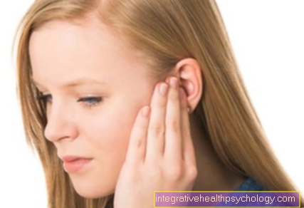
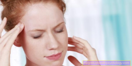
Pain in the tear ducts
Pain is one of the five signs of inflammation. Inflammation can also make itself felt in the tear ducts. Usually there is also swelling, reddening and overheating. Sometimes blocked tear ducts can also hurt. Tearstones (dacriolythes) can also irritate the tear ducts and cause pain. Treatment for the pain should be based on the cause. This is best determined by a doctor.
Find out more about the topic: Pain in the eye
Fistula on the tear ducts
A fistula is a naturally non-existent connection between a hollow organ and another organ of the body or the surface of the body. It is mostly tubular. The entire tear duct can be described as a kind of hollow organ, so fistulas can also form here.
Such fistulas can be congenital or form again as a result of melting and remodeling of tissue, e.g. as part of an inflammation. If the fistula ends on the surface, it can resemble a pus-filled pimple. Fistulas that are permanently inflamed and causing problems should be removed. This is done surgically. The inflammation is also treated with antibiotics