Narrowing of the trachea
definition
A narrowing of the windpipe (tracheal stenosis) describes a reduction or narrowing of the windpipe (Trachea). The trachea connects the lung with the Larynx and enables the transport of air to be inhaled and exhaled. If there is a narrowing in the trachea, the airflow can be restricted so much that the patient does Shortness of breath to get.
causes
There are various possible reasons for a tracheal stenosis.
One reason may be that there is an injury or inflammation of the windpipe (Trachea) has taken place. During the healing of the inflammation or the injury is the physiological (natural) Tissue of the windpipe broken downbecause it no longer fulfills its function and is then replaced by scar tissue. This Scar tissue can lead to a narrowing of the windpipe (tracheal stenosis) because it does not grow as finely and in a limited way as the original tissue. It has become a stenosing (constricting) scar formed.
Tracheal stenosis can also develop in the wake of a longer lasting one artificial respiration develop. Here, too, the reason lies in the development of scar tissue, which is caused by the long irritation of the windpipe by the Tube (Breathing tube) is formed as a protective mechanism. A similar reaction mechanism can also arise after Incisions in the trachea show where the scarring interface heals.
The tracheal stenosis can also be caused by a Cancer be evoked. If e.g. a tumor in the throat, on the thyroid glandthyroid or presses on the trachea starting from the trachea (windpipe) or relocates it from the inside, then there is a tracheal stenosis.
In regions where Iodine deficiency people often have a compensatory enlarged thyroid gland - this appears on the neck as Goiter (Goiter). If this presses on the trachea from the outside, it can become narrowed.
A narrowing of the trachea is ultimately also given when a patient hits an object or foreign body swallowed and it slips into the trachea and becomes stuck there. By relocating the trachea, depending on the size and location of the object, a small to large part of the lungs can no longer be ventilated and the patient complains accordingly shortness of breath.
Diagnosis and symptoms
The diagnosis is made via a ENT doctor. If the presence of one is suspected Tracheal stenosis becomes a CT scan made from the larynx and trachea. Ultrasound can also be performed.
To get a precise insight into the interior of the trachea, a reflection of the trachea is recommended. This is done either under local anesthesia or in general anesthetic.
For more precise diagnosis and planning of the treatment can depending on the location of the Narrowing a surgeon and Pulmonologist (Pulmonologist) to be drawn.
Depending on the strength of the narrowing of the trachea, the Symptoms only at; only when burden or one in peace. If the narrowing leads to a reduction of about Three quarters leads, then those affected have Shortness of breath in stressful situations. However, if the narrowing of the trachea leads to a reduction in diameter of more than three quarters, the affected people often have shortness of breath even in resting situations. If the constriction is very severe, step Sounds on inhalation. One speaks of one inspiratory stridor. The sound that is like a hum is caused by the fact that the air at the Bottleneck in the Trachea can not flow unhindered, but it comes to turbulence.
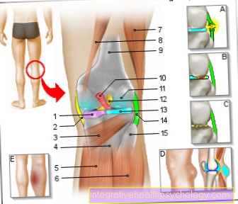
Illustration of the trachea

- Windpipe (approx. 20 cm) -
Trachea - Thyroid cartilage -
Cartilago thyroidea - Cricoid cartilage -
Cartilago cricoidea - Ring band -
Annular ligament - Tracheal cartilage -
Cartilago trachealis - Cover fabric - Tunica adventitia
- Trachea glands -
Glandulae tracheales - Mucous membrane - Tunica mucosa
- Membrane rear wall -
Pariesmembranaceus - Tracheal Muscle -
Tracheal muscle - Bronchiole - Bronchiolus
- Left lung -
Pulmo sinister - Left main bronchus -
Bronchus principalis sinister - Bifurcation of the windpipe -
Bifurcatio tracheae - Right main bronchus -
Bronchus principalis dexter - Right lung -
Pulmodexter
You can find an overview of all Dr-Gumpert images at: medical illustrations
OP and prophylaxis
A tracheal stenosis should be treated prophylactically not possible. However, patients with chronic complaints in the throat and throat area should consult a doctor at regular intervals for a possible einflammable Exclude the process and recognize it in good time.
Tracheal stenosis surgery is an operation that should be performed by well-trained doctors with several years of experience.
During the operation there is a stay of 1-2 weeks necessary. At the beginning, the patient will be using for several days Inhalation sprays treated. It contains drugs and substances (e.g. Cortisone) contain the Decongestion from the tissue in the operating area. In some cases there is also an prophylactic antibiotics Giving to combat possible pathogens of infections in the respiratory tract.
During the operation itself, the patient becomes a anesthesia offset. This anesthesia is maintained after the operation is completed and only the next day the patient is woken up again. The point is that the fresh surgical wounds spared become.
The trachea is exposed through a horizontal (from left to right or vice versa) incision on the neck above the Sternum. The neck muscles (i.a. Hyoid muscles) and the thyroid are pushed aside by the surgeon. The trachea is then exposed from all sides at the level of the narrowing and from the one behind it esophagus (Esophagus) detached.
Through cuts above and lengthways on the stenosis the trachea is opened. The tissue that caused the constriction is now away. The parts of the trachea that were above and below the stenosis are no longer connected to one another. Only that Breathing tube now allows the air to be transported into the lung.
The two ends of the trachea are now back to each other pulled up and sewn.
Last will water in the exposed space around the Trachea filled and air pumped. If no air escapes at the seam, the seam is tight. Then the water can be pumped out, the muscles pushed back and the neck closed again. Since it always closes Secondary bleeding can come Drainages (small plastic tubes) placed in the wound to allow any blood that has collected there to drain.
After the operation remains a scar back on the neck, but it is not necessarily visible. Within the first few days Pain around the surgical site occur. But in the long term no lasting discomfort stay behind.
Mostly one will be for the first few days after surgery antibiotic and decongestant drugs prescribed to the Healing process to support them as well as possible.
The patient can up to a quarter of a year be on sick leave and should take care of yourself.
Tracheal stenosis in the child
A innate Tracheal stenosis is coming very rare in front. If it occurs, however, then usually in connection with other deformities and malformations in the area of the Esophagus (Esophagus), further parts of the Respiratory tract (Respiratory tract) and am skeleton of the child. Depending on the extent and location of the stenosis, the severity of the symptoms varies.
With stenoses, the one rather short distance can be operated on. However, should the congenital stenosis reach the trachea (windpipe) via a longer distance can narrow a Tracheoplasty be performed.
Usually enclose horseshoe-shaped cartilage braces the trachea (windpipe) from the ventral (front). From the dorsal (back) the windpipe is from one connective tissue membrane delimited from the esophagus (esophagus). In children with a congenital narrowing of the trachea, some of these are cartilage braces very often malformed and enclose the entire trachea (Trachea) as a Cartilage ring. As a result, it is severely narrowed at these points.
At a Slide tracheoplasty ("Slide") is the windpipe (trachea) in the middle of the stenosis horizontal cut through: it is now divided into two parts upper and lower Part. Next, the sections of the trachea above and below the stenosis (narrowing) are cut open. Above the stenosis it becomes open at the front and below the stenosis it becomes open at the back: you can now look into the upper part from the front or into the lower part from the back into the windpipe. The two parts are now opened so that the part above the stenosis only has a front wall and the part below the stenosis only has a rear wall. In the next step, these two parts of the trachea are now pushed onto each otherso that the front wall and rear wall are on top of each other and sewn together can be.
That the trachea now a bit shorter is represents no problem that Lumens (Diameter) of the trachea is now but significantly enlarged, in that the cartilage clasps are bent up in the shape of a horseshoe and now two former small rings form a single large ring.
Depending on whether further malformations are diagnosed in the child, further interventions on the heart and lungs can be carried out in the course of the operation.