canine

Humans have a set of 32 teeth, almost all of which have different names. A distinction is made between incisors (Incisivi), Canines ( Canini), Premolars and molars (molars) from each other. Some people lack the ability to develop their wisdom teeth, also known technically as the figure eight. These people then only have 28 teeth in their dentition, but missing wisdom teeth do not mean any functional restriction
definition
In technical terms, the canine is referred to as the dens caninus, or often just as Caninus designated. ( Dens = Latin for tooth, Caninus= Latin for coming from the dog).
In predators, the canine tooth is enlarged and is used to tear prey, here it is also referred to as a fang or fang.
In human dentition, the canine is in the dental arch between the incisors and the premolars (these are the two front molars).
The tooth gets its name from the kink that the dental arch makes in its place. The incisors together with the canines form a semicircle, the four molars (premolars and molars) run after the canines in a straight line to the rear Tuber (The tuberosities are small bony elevations behind the last molars. They are very important for the hold of a full denture because they practically never atrophy. In a fully edentulous dentition, the tuberosity is of little functional importance.
In the literature, the tubers are also called Articular tuberosity designated.
development
Every human has four canines.
The human dentition is divided into quadrants. The first quadrant is lying in upper jaw and extends from the middle between the front incisors to the right rear to the last molar. The second quadrant is also in upper jaw and extends from the middle between the front incisors to the last molar on the left side. Analogous to this are the third and the fourth quadrant in the lower jaw. (The quadrant designation also applies to edentulous jaws, but the position of the teeth must then be considered.)
There is a canine tooth in each quadrant. Humans have both deciduous and permanent canines. The canines usually erupt first in the lower jaw, then in the upper jaw. In the second, permanent set of teeth, the front incisors break first, then the side incisors. In the lower jaw, the canine teeth first erupt and then the premolars and molars. In the upper jaw, on the other hand, the fourth tooth in the row of teeth (the front premolar) often comes first and only then does the canine break through.
In this way, there is often a lack of space in the upper jaw, a gap must first be opened (further) with the help of orthodontic treatment so that the canine has enough space and can grow into the oral cavity undisturbed. If orthodontic treatment is neglected in the early stages of tooth eruption, the growing canine shifts the entire row of teeth or grows completely outside of this row of teeth. Both possibilities require a subsequent one orthodontic treatment, both from an aesthetic and functional point of view.
The Eruption of the permanent canines takes place with about eleven years, in girls usually a little earlier than in boys.

I - right upper jaw -
1st quadrant (11-18)
II - left upper jaw -
2nd quadrant (21-28)
III - lower jaw left -
3rd quadrant (31-38)
IV - lower jaw right -
4th quadrant (41-48)
- 1. Incisor -
Dens incisivus I - 2nd incisor -
Dens incisivus II - Canine tooth -
Dens caninus - 1st molar tooth
Anterior tooth (premolar) -
Dens premoralis I. - 2nd anterior molar
Anterior tooth (premolar) -
Dens premoralis II - 1st molar tooth -
Dens molaris I - 2nd molar tooth -
Dens molaris II - Wisdom tooth (= 3rd molar) -
Dens molaris tertius
(Dens serotinus)
1st - 3rd are front teeth
(3 per quadrant)
4th - 8th are molars
(5 per quadrant)
You can find an overview of all Dr-Gumpert images at: medical illustrations
Look
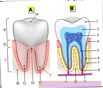
The Tooth crown of the canine Has no chewing surface but a Cusp tip with two cutting edges.
Looking at the canine of vestibular (from the outside, or from the inside of the lips or cheeks), that's how you see it Surface of the canine divided into two parts is. Both facets make one very shallow angles to each other. This middle edge is also the kink where the dental arch bends and runs backwards.
On the palatal Side in the upper jaw (the side facing the palate) and the lingual Side of the lower jaw (the side that points towards the tongue) can be seen two strong marginal ridges each mesial and distalwhich are in the middle together with the middle bar to form a strong Tubercle unite. Mesial always means towards the center of the dental arch; accordingly, distal is the surface facing away from the center of the dental arch.
The distal incisal edge of the canine is slightly longer than the mesial, and also forms a slightly flatter angle.
The Approximal surfaces are at the canines triangular. Approximal surfaces are the surfaces where two adjacent teeth touch.
In the course of life, the approximal surfaces become more flat and wider, the tip of the canine flattens out and is no longer quite as pointed. Both are perfectly normal Wear and tear which usually do not require treatment.
Canine tips are also the first to go away in patients with bruxism. They are just gnashed. (Bruxism is the technical term for crunch)
The root of the canine in the upper jaw is the longest in the entire jaw. In the upper jaw it can even reach almost to the paranasal sinuses. Canines have in almost all cases only one root canal. The The root itself is oval and inclined distally. The roots of the lower canine are shorter than those of the canines in the upper jaw.
Functions
The canines play one very important role in chewing.
When the rows of teeth are closed, the molars of the lower and upper jaw touch each other. If the lower jaw is now pushed to the left, as inevitably happens when chewing, the contact between the molars disappears. The tip of the canine slides along its counterpart in the other jaw, creating a small gap between the molars of the upper and lower jaw. The Canines ensure that the upper and lower jaws cannot remain pressed together with the molars when the lower jaw is moved to the right or left. So that will prevents excessive forces acting on the molars when chewing can. The consequence of constant excessive force on the teeth are namely Tooth loosening and that is more than undesirable. The whole prying out of the jaw by the canines is called Anterior canine guidance. This leadership is very important and should be maintained whenever possible. The tips of the canines become flatter in the course of life, but this is normal and does not require treatment. Under certain conditions, the tip of the canine can also be trimmed as part of an orthodontic treatment if the tooth is to replace a missing anterior tooth for aesthetic reasons. Functionally, an attempt should be made in this case to restore the above-mentioned anterior canine guidance at least in part with a premolar by grinding in the tooth.
When making crowns or bridges that also include the canine, the dental technician should absolutely dMake sure that the anterior canine guidance is retained, or is restored.
In the case of full dentures, the experts do not yet agree on whether an anterior canine guidance should be aimed for or not.
The Canine is very important for aesthetics and function, therefore, in the event of an extraction during orthodontic treatment, a premolar should preferably be removed to make room. Visually, premolars cannot be converted into canines very well, whereas canines can well be regarded as anterior teeth after grinding the tip.
Diseases
Retained canines in the upper jaw are relatively common. Due to the late eruption, the canine hardly has any space and then appears completely outside the dental arch, from where it can be removed with the help of Brackets and one stuck Braces must be placed back into the arch. The bracket is glued to the crown of the tooth and the tooth is pulled into the gap created by means of bands.
Sometimes the tooth is also lying so across the jaw that he only surgically removed become can. Sometimes the attempt becomes one Tooth transplant started and as part of the operation, the tooth was also immediately transplanted to where it belongs, but this is only possible in the rarest of cases.
A Failure to plant the canines is not very common and almost never occurs. A deciduous canine that does not want to fall out even in teenage or adulthood is therefore a good indication of dislocated teeth. Deciduous teeth are resorbed by the permanent teeth at the root and then fall out at some point due to the lack of support. If the following tooth is missing, it also happens no resorption and the milk tooth is preserved. Baby teeth are smaller and more bluish in color than permanent teeth.
The canines of the upper jaw are also called "Eye tooth" designated. The name comes from the fact that its root reaches almost to the bony orbit (the bony eye socket). Inflammations at the root tip are therefore not infrequently manifested Swelling and Redness below the eye. Purulent abscesses that form in the context of untreated root inflammation can therefore break into the eye socket or massively obstruct the view because the eye swells.
The dentist carefully opens the abscess and allows the pus to drain away. The swelling of the eye should then go away immediately.