Enchondroma
All information given here is only of a general nature, tumor therapy always belongs in the hands of an experienced oncologist!
Synonyms
Central (osteo-) chondroma, chondroma
multiple enchondromatosis: generalized enchondromatosis, dyschondroplasia, skeletal chondromatosis, Ollier's disease, Mafucci syndrome, chondroma within a bone, chondroblastoma.
English: enchondroma

definition
Under a Enchondroma one understands a benign Bone tumor cartilaginous tissue of origin (chondroma) within a bone.
An enchondroma is the most common tumor that appears within the small long bones hand and foot, or on pool, or is to be found in large tubular bones. In these areas in particular, this tumor, which consists of mature cartilage cells, grows into the soft parts of a bone (= medullary cavity) of the respective bone. Enchondromas are usually benign, but malignant degeneration can be observed in around 20% of all cases. The likelihood of malignant enchondroma is increased if the tumor is close to the trunk of the body.
There are six types multiple enchondromas. All types are considered developmental and calcification disorders, which usually manifest themselves in early childhood. They occur most frequently in the form of deformities or as indolent swellings in the hands and feet and thus represent tumor-like tissue malformations (tumor-like lesions).
Special forms:
M. Ollier (Ollier's disease): The tubular and flat bones are affected by chondromas on one side. In about 30% of all cases there is a malignant endurance, the so-called Chondrosarcomas.
Mafucci Syndrome: multiple enchondromas occur in combination with multiple cavernous ones Hemangiomas (Blood sponge) on the skin and internal organs.
If there is a degeneration, the rate of which is to be set as quite high in this area, one speaks of one secondary chondrosarcoma.
Summary
Under a Enchondroma one understands Chondroma cartilaginous tissue of origin within a bone. An enchondroma is the most common tumor that can be found within the small tubular bones of the hand and foot, or on the pelvis or in large tubular bones. In these areas in particular, this tumor, which consists of mature cartilage cells, grows into the soft parts of a bone (= medullary cavity) of the respective bone. For common are enchondromas benign, however, malignant degeneration can be observed in around 20% of all cases. The likelihood of a malignant enchondroma is increased if the tumor is close to the trunk.
Symptoms
Many make themselves in hand Enchondromas by gradually occurring, slowly increasing swelling in the area of the bone leading from the Enchondroma is affected, noticeable.
Not infrequent, however Enchondromas Also discovered during an X-ray examination of the hand, which was carried out for completely different reasons (e.g. after an accident).
metastasis
Enchondromas can become malignant with a probability of 20%. Entrapment particularly affects those patients in whom enchondromas already occur more frequently (= multiple occurrences)
In cases in which enchondromas have already colonized the larger tubular bones or the pelvis, the probability of malignant degeneration is increased.
In summary, it can be said that degenerations are more likely the more they occur, or when the occurrence has already spread to areas outside the small tubular bones on the hand and foot.
Enchondromas do not metastasize.
diagnosis
As a rule, X-ray diagnostics are used for diagnosis. As a rule, central, oval-shaped osteolysis then becomes visible. In addition, in cases of spontaneous fractures, the cortex (= outer, hard layer of the bone).
X-ray diagnostics alone often do not allow a reliable diagnosis. In the case of an enchondroma, characteristic changes can be seen on the X-ray, but such changes can also be seen in other (possibly malignant) tumors.
Ultimately, only that provides information histological examination of the tumor tissue. Such histological examinations are generally carried out routinely during the surgical removal.
Imaging
Imaging must be done to assess an enchondroma. The most common, cheapest, and easiest to perform imaging is a X-ray image. Accordingly, if an enchondroma is suspected, an X-ray image is usually taken first. Often this is sufficient for an assessment.
If this is not the case, a MRI (Magnetic resonance imaging). Computed tomography examinations are also possible, but are associated with a significantly higher radiation exposure than X-rays or MRIs.
Complications
Every operation carries a certain risk. That can never be ruled out from the outset.
So can also in the context of this operation Disorders of wound healing or in rare, severe cases, severe bone cavity infection occurs. These symptoms are then treated with antibiotics, and reoperation is rarely necessary.
Since enchondromas are localized differently, different difficulties can arise in the course of an operation, depending on the individual situation. For example, injuries in the area of tendons and / or nerves cannot be ruled out.
Especially if the enchondroma is located near the joint, you can Mobility disorders occur, which can, however, be counteracted by appropriate therapeutic measures.
Sudeck's syndrome, the Sudeck's disease (CRPS) especially after fractures and Soft tissue injuries, poor immobilization. Multiple repositioning or after traumatizing surgical techniques can appear. The result is a painful mobility disorder. To find out more about this topic, please click on the link.
During planned operations, you should stop taking blood-thinning drugs (e.g. aspirin, ASS, Marcumar, etc.) must be discontinued about 10 - 12 days before the planned operation in agreement with your treating doctor.
Consultation with the attending physician is so important because some patients continue to show reduced blood clotting even after discontinuing the preparation and this may have to be treated with heparin before the operation.In many patients it has been shown that the use of the above drugs can be interrupted for a few weeks. Only your attending physician can understand which of the two groups you belong to. !
Please also note that unconscious ingestion of aspirin in the event of toothache and / or headache in the period of 10. Should be omitted 12 days before the operation. In severe cases, please choose pain relievers that do not have any blood-thinning properties. An example is at this point Paracetamol mentioned.
How is the enchondroma treated?
Depending on the individual appearance, there are different therapeutic measures and options.
Enchondromas iIn the area of the small tubular bones, if there are no symptoms and there is no risk of fracture, usually no treatment at all. If this is not guaranteed, curettages (= surgical scraping of the affected areas) and autologous cancellous bone fillings can be initiated.
If the enchondromas relate to the area of the long tubular bones and the trunk skeleton, the type of enchondroma must first be analyzed, lying calcifying enchondromas before, no therapy is sought - in the case of no symptoms. The patient observes himself and comes for an x-ray check-up once a year. Are others lying Enchondroma-forms In this area before, a curettage and a cancellous plastic are usually performed.
If there is an urgent suspicion of malignancy, an immediate one is usually found Enchondroma removal instead of.
As part of the enchondroma removal, an incision is first made in the tumor area through the skin and into the area above bone lying structures. Finally, the cortex (= outer, hard layer of the bone) is opened in order to scrape the tumor through this access. In order to be sure that the last remnants of the resident tumor could be removed, the tumor area is rinsed after scraping.
Since the tumor was within the bone, it is possible that after the surgical removal of the enchondroma (e.g. in the hand) a very large "hole" remains within the bone. In such cases, bone tissue is removed from another point in order to reinsert it in the former tumor area and thus fill the hole that has formed.
As a rule, this transplantation takes place very rarely, since it has now been scientifically proven that in many cases the body can fill the cavity in the bone with independently newly formed bone tissue.
So it can be said that a transplant will only be carried out if the bone is through the Enchondroma has already been severely damaged and its stability seems to be greatly reduced.
In the case of multiple enchondromas (enchondromatoses), limb shortening may be necessary. These can then be compensated for with equipment or by means of lengthening osteotomies.
Aftercare
The operation is followed by immobilization, the duration of which depends on the extent of the enchondroma. The following rule of thumb applies:
The more extensive the enchondroma, the longer the period of immobilization after the operation.
Scars appear on the operated site, but these are barely noticeable after 1 - 2 years. The Scars are particularly sensitive in the first few days and weeks after the operation. Sensory disturbances can occur particularly during operations on the hand, which has a very high density of sensory nerves.
Postoperatively, pain and swelling can occur for weeks or months. This is especially the case when the hand is used again in everyday use. The scar in particular can then also be thickened and reddened. These symptoms should completely subside within the first year after the operation.
Important NOTE:
The hand can usually be used again in everyday life about six weeks after the operation. However, care must be taken to avoid overstressing. For this reason, certain sports activities should be avoided for about four to five months after the operation. However, this time can be extended if the enchondroma has already severely affected the bone (thinning of the cortex, ...). In such cases, sports leave of around 6 months is conceivable.
Even if the hand can be used again in everyday life about six weeks after the operation, the hand should be gradually brought back to these activities. Too fast a transition can lead to pain and overload, which in turn has a negative effect on healing.
After the bandages have been removed, the operated hand can be regularly bathed in lukewarm water for about three times for five minutes over a period of about two months. Light movements without great effort or strain should be performed regularly.
Physiotherapy:
Patients who are able to move their hands independently without stress usually do not need physiotherapy. It is reserved for those patients whose area of operation shows insufficient mobility.
In general, movements - regardless of whether they are performed independently or as part of physiotherapy - should never lead to pain or swelling.
Enchondroma on the finger
Enchondromas mainly occur in the area long tubular bones on, these include the finger. So this is the most common localization from the cartilage tumors. Enchondromas are less common in the thigh, upper arm, foot and pelvis.
They usually grow slowly and in most cases do not cause any discomfort. Therefore, if at all, they are often only noticed as a chance finding when taking an X-ray. They can only be seen from the outside as a slowly increasing swelling as they grow in size. Enchondromas in the area of the fingers can occasionally become too Pain to lead. In addition, they can lead to a reduction in the stability of the bones due to the slow displacement of the bone tissue. This can lead to the bone breaking under normal stress. One speaks of one pathological fracture.
In spite of the rare degeneration, if an enchondroma is detected, regular check-ups by means of an X-ray should be carried out in order to recognize rapid growth in size and markers that may indicate a degeneration should be recognized in good time. If an X-ray cannot safely rule out that the lesion is malignant, other imaging methods such as magnetic resonance imaging and computed tomography are used; in case of doubt, the tumor must be surgically removed. Then a tissue sample of the tumor is sent to a pathological institute. In most cases, it is possible to distinguish between a benign and a malignant lesion under the microscope.
However, an enchondroma can not only be surgically removed if a degeneration is suspected. Surgical removal can also be useful if the person concerned repeatedly complains of complaints - for example pain - or fractures occur. Here, an incision is made over the tumor tissue, the tumor is peeled out and the tumor cavity is then thoroughly rinsed to remove residual tumor. Such an operation can usually be carried out under local anesthesia, so no real anesthesia is usually necessary.
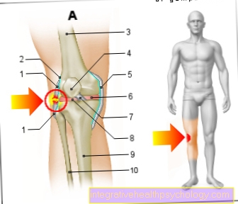
Enchondroma on the knee
Enchondromas are cartilage-made tumors that are benign in most cases. Most often they occur in the area of the fingers. They are less common in the area of Thigh and also des Knees to find.
Often times, enchondromas are complete asymptomaticso make no complaints. However, if there is an increase in size, mobility can be impaired. In particular, if an enchondroma grows in the knee joint, the Knee flexion difficult or be painful. In such cases, surgical removal should be sought. Imaging should be done beforehand. As a rule, an X-ray is taken first. If the findings cannot be adequately assessed here, a magnetic resonance examination can also help.
It is true that enchondromas that are close to the trunk, i.e. near the center of the body, have a higher risk of degeneration than those that are in the periphery. This means that with an enchondroma of the knee there is a higher risk of degeneration than with an enchondroma of the fingers. It is therefore usually advisable to either have an enchondroma of the knee checked closely or to have it surgically removed. Depending on the size of the tumor, the tumor cavity must be filled with bone after removal. You can use your own bone (e.g. from the iliac crest) or someone else's bone.


.jpg)