rectum
Structure of the rectum
The colon makes an S-shaped bend. This section is called the sigmoideum. It is the last link between the large intestine and the final rectum. The rectum is also known as the rectum.
Above all, it is a reservoir and stores prepared stool that is intended to be eliminated. The rectum begins approximately at the level of the sacrum.
The rectum has a length of approx. 15-20 cm. It ends in the anus, which is formed by the sphincter muscles as well as the perineal muscles. These sphincters hold back the bowel movement and thus ensure sufficient continence. The inside of the rectum is criss-crossed with a plexus of veins. If this vascular system bulges, the well-known hemorrhoids occur. Such haemorrhoids can develop, especially with solid stool or with increased pressures during defecation.
There are several stages of hemorrhoids. Bulging vessels always present the risk of injury. If this happens, one speaks of hemorrhoid bleeding, which can be not insignificant. Venous plexus sacs can be treated with a variety of ointments, or surgery can be performed.
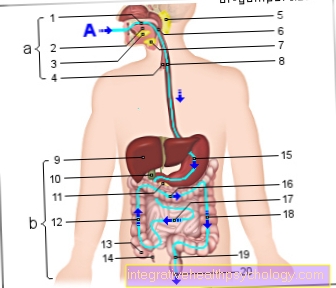
Figure digestive tract

Digestive tract
A. - Food route
a - digestive organs
in the head and neck
(upper part of the digestive tract)
b - digestive organs
in the body cavity
(lower part of the digestive tract)
- Oral cavity - Cavitas oris
- Tongue - Lingua
- Sublingual salivary gland -
Sublingual gland - Trachea - Trachea
- Parotid gland -
Parotid gland - Throat - Pharynx
- Mandibular salivary gland -
Submandibular gland - Esophagus - Esophagus
- Liver - Hepar
- Gallbladder - Vesica biliaris
- Pancreas - Pancreas
- Colon, ascending part -
Ascending colon - Appendix - Caecum
- Appendix -
Appendix vermiformis - Stomach - Guest
- Large intestine, transverse part -
Transverse colon - Small intestine - Intestine tenue
- Colon, descending part -
Descending colon - Rectum - Rectum
- Nach - anus
You can find an overview of all Dr-Gumpert images at: medical illustrations
In diseases of the intestine that require surgical removal of sections of the intestine, it is important that a large part of the rectum is preserved. Otherwise there is a great risk of one Incontinence. Does it come to blood deposits in the stool or so-called in patients Tarry stool, should definitely pass the bowel through a reflection (Colonoscopy) to be examined.
A so-called digital rectal examination should be done in any case if blood is detected in the stool.
Here the rectal wall can be felt, constrictions can be found and it can also be checked whether the ampoule of the rectum is filled with stool and whether this is blood-free or if there is blood.
If the infestation is pronounced, the digital rectal examination can already suspect a Rectal cancer which can be noticeable in a pronounced constriction.
In addition to the digital rectal examination, one should definitely have one if there is any suspicion Rectoscopy carry out. This is a colonoscopy in which only the rectum is seen. To carry out this procedure, much less effort and preparation is required than with a "large" colonoscopy.
Most of the time, shortly before the procedure, the patient only gets a purgative suppository to empty the rectum and thus allow a corresponding insight. A rigid instrument is then inserted into the anus and the rectum is inspected while advancing.
Figure large intestine

- Colon, ascending part -
Ascending colon - Appendix - Caecum
- Appendix -
Appendix vermiformis - Right colon bend -
Flexura coli dextra - Large intestine, transverse part -
Transverse colon - Left Colon Bend -
Flexura coli sinistra - Colon, descending part -
Descending colon - Large intestine, s-shaped part -
Sigmoid colon - Rectum - Rectum
- Bulges of the
Colon Wall -
Haustra coli - Liver - Hepar
- Stomach - Guest
- Spleen - Sink
- Gallbladder -
Vesica biliaris - Small intestine -
Intestine tenue - Esophagus -
Esophagus
You can find an overview of all Dr-Gumpert images at: medical illustrations