Vascular supply to the heart
Synonyms in a broader sense
- Coronary arteries
- Angina pectoris
General
When speaking of vascular supply (vascular supply to the heart / coronary arteries), one must first differentiate between arteries, veins and lymphatic vessels. While arteries bring the oxygen-rich blood to the respective target organ, the oxygen-poor blood is transported back to the heart via veins after perfusion of the organ. Lymph vessels begin blindly at every organ and carry intracellular fluid via larger lymph vessels into the superior vena cava (superior vena cava).
Many vascular supplies can be deduced if one knows the embryonic development of humans, since, to put it simply, there are different zones that are later supplied by the same vessels. With knowledge of the embryonic development one can derive many vascular supplies.

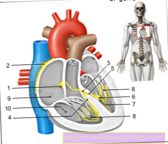
- Right atrial -
Atrium dextrum - Right ventricle -
Ventriculus dexter - Left atrium -
Atrium sinistrum - Left ventricle -
Ventriculus sinister - Aortic arch - Arcus aortae
- Superior vena cava -
Superior vena cava - Lower vena cava -
Inferior vena cava - Pulmonary artery trunk -
Pulmonary trunk - Left pulmonary veins -
Venae pulmonales sinastrae - Right pulmonary veins -
Venae pulmonales dextrae - Mitral valve - Valva mitralis
- Tricuspid valve -
Tricuspid valva - Chamber partition -
Interventricular septum - Aortic valve - Valva aortae
- Papillary muscle -
Papillary muscle
You can find an overview of all Dr-Gumpert images at: medical illustrations
Systematics of the lymph vessels
In all parts of the body, the smallest lymph capillaries begin blindly. This means that there intracellular fluid flows into the smallest capillary. Part of the lymphatic system are also the lymphatic organs (e.g. spleen and Almonds), in which the differentiation of T lymphocytes takes place. These lymph capillaries then close local lymph nodes together, where the lymph fluid collects and then continues in larger lymph vessels to the Milk breast duct (Thoracic duct), the body's largest lymphatic vessel. Through manual therapy (targeted massage), the outflow of the lymph can be increased and thus Lymphedema (Water accumulation) must be eliminated. In addition, lymphedema can also go through Compresses or Medication be treated. Another interesting aspect of the Lymphatic system are the lymph nodes associated with cancer (Metastasis). An example of this is the lymph drainage from the breast, which runs through the lymph nodes in the armpits. There at Breast cancer Cancer cells reach these lymph nodes via the lymph, metastases often form here, so that the lymph nodes also have to be removed (vascular supply to the heart).
Systematic blood vessels

The central organ of the blood supply is the heart (Vascular supply heart) It pumps deoxygenated blood to enrich the oxygen lung and then in the Body circulation. The largest artery in the body is that artery (aorta). All of the vessels that ensure the arterial supply of the various tissues in the body emanate from it. Of the venous return of blood takes place depending on the position of the tissue via the superior or inferior vena cava (Vena cava superior / inferior). There is also a kind of diversion system that runs parallel to the vena cava in the chest and abdomen. On the one hand, this is the one Azygos vein, on the other hand the hemiazygous vein. Both are connected to each other at the level of the heart and finally flow into the superior vena cava. Another special feature of the blood supply is that Portal vein system of the liver The venous blood of all unpaired abdominal organs (everything except genital organs and Kidneys) flows over the Portal vein (Porta vein) in the liver. There the nutrients in the blood are metabolized in the liver. The blood then passes through the liver veins (Hepatic veins) into the inferior vena cava (Inferior vena cava) (Vascular supply to the heart).
Vascular supply to the heart
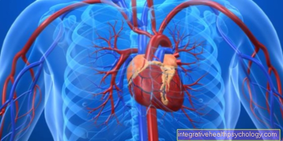
The heart (Cor) is a muscular hollow organ that plays the central role in the vascular supply of the body (Vascular supply heart). As a pump, it transports oxygen-poor blood to the lungs (Pulmo), where the blood is enriched with oxygen. The heart then pumps the oxygen-rich blood back into the body's circulation.
The heart itself becomes through two Coronary arteries (Coronary arteries) provided. The right side of the heart is formed by the right coronary artery (right coronary artery, dextra coronary artery), while the left side of the heart is supplied by the left coronary artery (left coronary artery, left coronary artery) is supplied. Both coronary arteries leave the main artery (aorta) immediately after it emerges from the heart. This ensures that particularly oxygen-rich blood supplies the heart. The right coronary artery supplies not only the right part of the heart but also the front third of the heart septum (Septum cardiale) and the Sinus node as well as the AV node (Atrioventricular node). (The left coronary artery divides at the beginning into the Ramus circumflexus and the Ramus interventricularis anterior).
Depending on which of the two coronary arteries takes over the main supply of the heart, one speaks of Left supplier type (if the main supply is from the left coronary artery), from Legal provider type (if the main supply is through the right coronary artery) and at Normal supply of the intermediate type.
The venous blood of the heart overflows three main branches, the Vena cardia parva, media and magna in the Coronary vein sinus (Coronary sinus). The coronary vein sinus joins the upper and lower vena cava (vena cava superior et inferior) in the right atrium (Atrium dextrum) (Vascular supply to the heart).
Vascular supply of the pericardium
Of the Pericardium surrounds the heart and serves as a sliding space. The arterial supply (vascular supply to the heart) takes place via the Diaphragmatic sac artery (Pericardiac artery) from the internal thoracic artery (Internal thoracic artery) arises. The venous outflow takes place via the Diaphragmatic sac vein (Pericardiac vein) in the internal thoracic vein (Internal thoracic vein), which ultimately goes into the superior vena cava (Superior vena cava) opens (vascular supply to the heart).


.jpg)