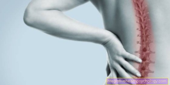
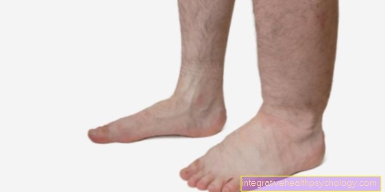
Joint effusion in the knee
introduction
When there is a joint effusion in the knee, fluid collects in the knee joint. These are often Synovial fluidthat of the Synovial membrane (Synovia) is produced in excessive quantities.
However, blood (Hemarthrosis) or pus (Pyarthrosis) accumulate in the knee.
The affected patients often complain of pain and restricted mobility of the knee.
A joint effusion is not an independent disease, but a symptom that can occur as part of injuries, infections or other diseases of the knee joint.
Hence the therapy is usually based on that Underlying disease, the cause of the effusion.
Also read more on the topic: Bruise in the knee

root cause
The causes of a joint effusion in the knee can be mechanical or inflammatory in nature.
An overload of the knee joint due to exercise is a possible mechanical cause, particularly in young patients.
In older people, on the other hand, joint effusion is often caused by wear and tear on the joint (osteoarthritis). In many cases, however, a joint effusion in the knee is a symptom of an injury, for example after a fall or an accident. Joint effusion can occur with the following knee injuries:
- Damage to the meniscus
- Cruciate ligament tear
- Inner or outer ligament tear
- Fracture of the kneecap, the head of the tibia, or the femur
- Contusion, sprain, or twist (distortion)
- Even after an operation on the knee, an effusion may develop over a longer period of time.
You might also be interested in this topic: Cruciate ligament overstretched
Other mechanical causes can be gout crystals or bone tumors in the knee joint area.
While effusions caused by wear and tear or overload usually contain synovial fluid, after injuries it is not uncommon for a bloody joint effusion to occur.
In particular, rheumatic diseases and various other forms of arthritis come into question as inflammation-related causes.
But bacterial infections can also cause a joint effusion in the knee, in which case it is often a purulent effusion.
Symptoms
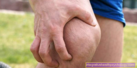
A joint effusion in the knee shows up externally as a swelling of the knee joint, through which the contours of the joint look like.
Read more on the topic: Joint swelling of the knee
As a rule, the mobility of the joint is limited, so that some patients can hardly bend or straighten the knee.
In addition, those affected usually complain of pain in the joint.
If the cause of the joint effusion is inflammatory, the typical symptoms of inflammation can also occur: The knee is then reddened and overheated.
You might also be interested in this topic: Lump on knee
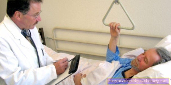
Appointment with a knee specialist?
I would be happy to advise you!
Who am I?
My name is I am a specialist in orthopedics and the founder of .
Various television programs and print media report regularly about my work. On HR television you can see me every 6 weeks live on "Hallo Hessen".
But now enough is indicated ;-)
The knee joint is one of the joints with the greatest stress.
Therefore, the treatment of the knee joint (e.g. meniscus tear, cartilage damage, cruciate ligament damage, runner's knee, etc.) requires a lot of experience.
I treat a wide variety of knee diseases in a conservative way.
The aim of any treatment is treatment without surgery.
Which therapy achieves the best results in the long term can only be determined after looking at all of the information (Examination, X-ray, ultrasound, MRI, etc.) be assessed.
You can find me in:
- - your orthopedic surgeon
14
Directly to the online appointment arrangement
Unfortunately, it is currently only possible to make an appointment with private health insurers. I hope for your understanding!
Further information about myself can be found at
Diagnosis

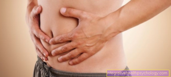
The diagnosis is based on a detailed questioning of the patient (anamnesis), through which the doctor receives information about possible causes of the complaints.
The subsequent physical examination plays an equally important role. The mobility of the joint is checked and various function tests are used to search for indications of an injury to the ligaments or menisci as a possible cause of the effusion.
A larger joint effusion in the knee can be diagnosed with certainty by the so-called "phenomenon of the dancing patella": The doctor spreads the synovial fluid above and below the knee joint and then presses the kneecap (patella) against the thigh with the index finger. If he feels a resilient resistance, this indicates a joint effusion, as the kneecap "floats" on the excessively existing synovial fluid.
Smaller effusions of a few milliliters can only be detected by the so-called "bulge sign": If you press on the side below the kneecap, a bulge becomes visible on the other side, which spreads as a small wave when you tap gently.
In many cases it makes sense to use additional imaging techniques. The fluid in the joint is clearly visible in the ultrasound, X-ray and MRI of the knee. In addition, the MRI images often provide information about the cause of the joint effusion, such as an injury.
If the cause of the joint effusion in the knee is still unclear, a knee puncture is usually performed. On the one hand, this relieves the joint by reducing the amount of fluid.
On the other hand, the biochemical examination of the liquid obtained can provide information about the cause. In the case of an inflammatory joint effusion, for example, a large number of white blood cells and protein can be found in the synovial fluid, whereas bacteria can be detected in the case of an infection.
Small crystals in the liquid, however, indicate a gout disease as the cause.
MRI and X-ray
To display the fluid in the knee, it is usually enough Ultrasound of the knee out.
Imaging procedures, like that MRI of the knee joint and X-rays also show the joint effusion well. However, they are more likely to be used when it is unclear where the joint effusion is coming from.
The roentgen of the knee joint is particularly suitable for depicting the skeletal structure and thus the structure of the knee joint.
Broken bones or splinters caused by accidents or trauma can be made recognizable. Also can Cartilage damage in the knee jointe.g. destroyed or worn cartilage like him at Knee osteoarthritis occurs.
In addition, osteoarthritis (Joint disease caused by damaged cartilage) noticeable through a narrowed joint space, which is also recorded in an X-ray.
The x-ray of the knee is usually made while standing, so that the joint is shown under the greatest and also natural load. If ligament structures, soft tissues in the knee joint and surrounding soft tissues or damage to the menisci are the reason for the joint effusion in the knee, this can be remedied with the MRI represent well.
It allows a three-dimensional representation of the knee joint and is suitable for detecting defects in the bone structure at an early stage. The MRT thus has an advantage over the X-ray, but it is also more costly and time-consuming.
However, MRI is also more of a selective procedure that is used for specific questions.
therapy
The treatment of a joint effusion in the knee depends initially on the underlying disease. If the cause is eliminated, the effusion often disappears as well.
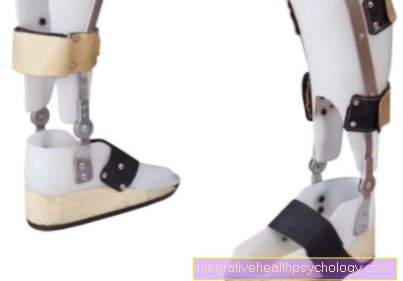
Basically, the affected joint should be protected, for example using a splint, and elevated. Cooling compresses have a slightly decongestant and pain-relieving effect.
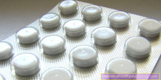
If the pain is severe, supportive anti-inflammatory pain relievers (NSAIDs) such as ibuprofen or diclofenac can be taken.
If the effusion is very large, liquid can be drawn off by a knee puncture, which relieves the joint.
homeopathy
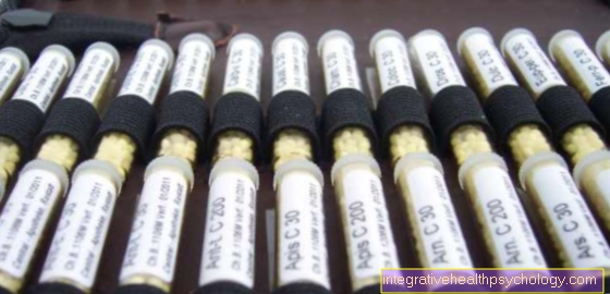
In homeopathy, potentized animal, herbal and mineral ingredients are used to treat diseases.
Also at Knee effusion homeopathic medicines can be used.
Is it a joint effusion caused by trauma (Accident, fall), low-potency drugs are selected and taken at shorter intervals (e.g. three times a day, in some acute cases every 30-60 minutes). This is how you take it three times a day for an acute joint effusion Bryonia alba (white bryony) until the swelling has subsided.
Accompanying is recommended Rhus toxicodendron (Oak-leaved poison oak) to take. This is the recommendation for knee joint effusions caused by accidents, falls and minor sports injuries. In the case of chronic diseases, drugs with a higher potency are usually chosen, but they do not have to be taken as often. If, for example, a joint effusion as a result of a chronic disease such as inflammatory osteoarthritis is to be treated, it is recommended to take it again Bryonia alba. In addition, it is recommended Apis mellifica (Western honey bee) to be administered. This should be done for about 10 days. If there is no improvement, it is always advisable to consult a doctor who will identify and treat any injured knee structures.
Dotting
The puncture of the knee joint can be a therapeutic and / or differential diagnostic means. During the puncture, the fluid is removed from the joint with a hollow needle (Cannula) pulled out, which firstly ensures pressure relief (therapy) and secondly serves to be able to examine the fluid from the knee (Differential diagnosis).
This liquid is then used to find the cause, as the effusion can be of different consistencies and colors.
There may also be bacteria in the liquid, which can be identified by examining the liquid.
Effusion fluid can be bloody if it is caused by trauma (often with sports injuries) is caused. It can also be purulent (fibrinous) if the disease in the joint is an infection. However, it can also be serous if the cause is a mechanical overload.
A puncture should only be performed if the joint is severely swollen or if there is evidence of joint effusion. If the effusion is caused by a chronic disease such as osteoarthritis, the underlying disease should also be treated, as it is very likely that an effusion will occur again.
Read more about this at: Knee puncture
forecast
If the cause is treated successfully, the joint effusion in the knee will often go away on its own after a while.
A chronic effusion can occur, however, when the synovial membrane constantly produces too much synovial fluid due to permanent irritation.
This can be for example at
- arthrosis
- arthritis
- or an untreated infection.
In some cases, a so-called Baker's cyst forms as a result of chronic joint effusion in the knee.
This is understood to be the bulging of a bursa into which the excessively produced synovial fluid drains. This is then noticeable as a palpable lump in the back of the knee that causes a feeling of pressure when the joint is bent. If the Baker's cyst causes severe discomfort, it may have to be surgically removed.
Duration
The duration of a knee joint effusion depends on whether the effusion is the result of an acute or a chronic disorder.
In addition, the duration depends on how serious the injury or illness is.
Is it a minor knee injury with a minor KneesswellingAs often happens in sports, cooling, raising and resting the leg can reduce the swelling or effusion within a few days.
If the injury is severe and requires surgery, it can take a few weeks or months to heal. The joint effusion was caused by a chronic Inflammation in the knee caused, this must be treated so that the fluid regresses.
This too can take days to weeks. With a Puncture of the knee joint the liquid is removed immediately. However, if the cause of the effusion is a chronic disease, a renewed effusion can be expected if the cause is not treated.