Function of the ovaries
Synonyms
Ovary, ovaries (pl.), Ovary, ovary, oophoron
English: ovary
function

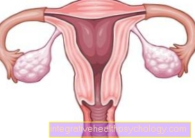
The ovaries are the woman's reproductive organs. On the one hand, the egg cells mature and are released into the fallopian tubes. On the other hand, the ovary is a production site for hormones (Estrogens, progestins).
These processes are controlled by the pituitary gland (pituitary gland), which releases hormones in a certain time schedule (secreted) and thus directs the ovarian cycle. These hormones are the pituitary gonadotropins FSH (= follicle-stimulating hormone) and LH (luteinizing hormone).
Illustration ovaries

- Ovary -
Ovary - Basic tissue of the ovary -
Stroma ovarii - Mature vesicle follicles -
Folliculus ovaricus tertiarius - Corpus luteum -
Corpus luteum - Uterine cavity -
Cavitas uteri - Cervix -
Ostium uteri - Ovarian ligament -
Ligamentum ovarii proprium - Fringed funnel of the fallopian tube -
Infundibulum tubae uterinae - Fallopian tubes -
Tuba uterina - Ovarian artery -
Ovarian artery
You can find an overview of all Dr-Gumpert images at: medical illustrations
The follicle in the Ovaries the woman are all together before the birth educated. No new follicles appear after birth.
At birth, women have 1 to 2 million follicles in both ovaries. However, these follicles are not yet mature. They are in a kind of dormant state for 12 to 50 years. In this resting stage, germ cell division is halted. The follicles are small and are called primordial follicles. In the fetal and childhood phases, as well as later in fertile adulthood, some of these primordial follicles repeatedly mature into tertiary follicles via primary and secondary follicles due to factors that are not yet understood.
The follicles become larger, but germ cell division is still stopped. In this tertiary stage, however, the follicles all die in the fetal and childhood phases, because the children do not yet secrete the hormones that the tertiary follicles need for further maturation and germ cell division. This process of death is called atresia.
With the beginning of the pubertyAt puberty, women only have around 400,000 follicles. From this, as in childhood, primordial follicles mature into tertiary follicles. Most of them die, as in childhood. However, 10 to 20 of them manage to mature further in each cycle due to the hormonal influence of the pituitary gland, which takes on its function during puberty.
By gonadotropins (FSH) these selected 10-20 follicles are influenced, one also speaks here of a cohort, getting bigger and bigger. One follicle is particularly sensitive to the hormone FSH and is thereby stimulated more than the other follicles in its cohort. This leads to this selected (selected) Follicle becomes the largest of all. It is known as the dominant follicle. Within a week it grows three times as much (approx. 25mm) and has now grown into what is known as a mature follicle. Since this selected follicle is the most sensitive to the FSH hormone, there are more admission sites (Receptors) for the hormone, he gets more FSH than the other follicles in the cohort, so to speak. The other follicles are therefore insufficiently influenced and therefore all die (Atresia).
The cohort stimulated by the hormone FSH also always forms hormones, namely estrogens, at the time of further maturation. The dominant follicle produces most of it. These hormones are important as they stimulate the uterus and also the mammary gland. More precisely, this means that the mucous membrane in the uterus is stimulated to grow (proliferate) in order to respond to a potential pregnancy and implantation of the germ to be prepared.
When the mature follicle is very well developed, the amount of estrogens produced in the ovaries is so great that the pituitary gland is stimulated to secrete the gonadotropic hormone LH. This LH, in turn, has an effect on the ovaries. The rise in this hormone causes it to ovulate (ovulation) is coming. The mature follicle now continues the germ cell division (1st meiosis is ended and the second meiosis begins). The Egg cell dissolves from the follicle cells and certain enzymes break down the follicle wall and the organ capsule so that the egg cell and the fluid in the follicle can find a way towards it Fallopian tube (tuba uterina) can pave the way. The egg cell is then picked up by the fallopian tube. In the case of a fertilization the egg cell completes its 2nd meiosis.
The remnants of the follicle, i.e. follicle cells without an egg cell, develop into the so-called after ovulation Corpus luteum menstruationis. These cells convert something and now produce progestins such as, for example progesterone. This hormone has the task of maintaining a possible pregnancy and is formed for this very reason.
The greatest amount of progestin is formed on the 7th day after ovulation. In total, such a corpus luteum lasts 14 days if no fertilization takes place. Then the corpus luteum perishes (Luteolysis) and a white scar forms. The corpus luteum is now called Corpus luteum albicans designated. Gestagens are no longer produced, so that the pituitary gland is stimulated to release FSH again, so that a new cohort can then be recruited and the cycle starts all over again.
In the case of pregnancy, the corpus luteum persists for two months and is produced by an LH-like hormone (HCG), which is formed by the fertilized germ, further progestins and thus maintains the pregnancy. The corpus luteum, which during the pregnancy is known as the corpus luteum graviditatis.
The ovaries can also cause pain during pregnancy. Information on this topic can be found at Ovarian pain in pregnancy.

