Smooth musculature
definition
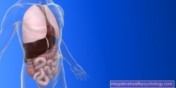
Smooth muscles are the type of muscles that are found in most of the human hollow organs and, due to their special structure, are able to work very effectively and independently without a high expenditure of energy.

properties
The smooth muscles got their name from the fact that they are derived from the other type of muscle, namely the muscles that are found primarily in the skeletal muscles and are known as striated muscles clearly differentiates. This is because these muscles show transverse stripes under polarizing light, which is caused by the regular arrangement of the proteins actin and myosin.
This one regular order in the smooth muscles is missing, the muscle cells appear homogeneous here even under polarizing light.
Building the smooth muscles
Typically, the smooth muscle cells are that too Myocytes to be named, spindle-shaped and have a diameter of about 5 to 8 µm. Of course, this depends on the state in which the cell is: in a contracted (contracted) muscle the cells are slightly thicker than in a flaccid one.
The The length of the muscle cells varies especially strong, not only because of the state of contraction, but also depending on the location the cell.
In the Blood vessels the cells are on average only 15 to 20 µm long, in other organs they can reach a length of 200 or 300 µm and in the uterus In a pregnant woman, muscle cells can even grow up to 600 µm in length through special adaptation processes.
Cell nuclei smooth muscles are usually also somewhat elongated and are typically located, just like most other cell organelles (endoplasmic reticulum, mitochondria, ribosomes, etc.), in the middle of the cell.
The Filaments actin and myosinAs mentioned above, these muscle cells are also present in high concentrations in the cytoplasm, but are subject to them not such a strict structure like in the striated muscle cells. Here they pull rather disordered more or less criss-cross through the muscle cell, within the cytoplasm at the so-called dense bodies ("Dense body", in German sometimes also known as compression points) and at the edge of the cell Anchoring plaques are attached.
This arrangement has the consequence that a single cell and thus also the whole muscle contract when it contracts contract more can than is the case with a striated muscle.
Every single cell is covered by a thin skin that Basal lamina, surround. Usually several cells are then arranged in groups, either very tightly packed or in the form of smaller bundles.
Figure muscle fiber

- Muscle fiber
of a skeletal muscle
Muscle fibra - Muscle fiber bundles -
Muscular Fasciculus - Epimysium (light blue) -
Connective tissue sheaths around groups
of muscle fiber bundles - Perimysium (yellow) -
Connective tissue sheaths
around muscle fiber bundles - Endomysium (green) -
Connective tissue between muscle fibers - Myofibrils (= muscle fibrils)
- Sarcomere (myofibril segment)
- Myosin threads
- Actin threads
- artery
- vein
- Muscle fascia
(= Muscle skin) - Fascia - Transition of muscle fibers
in tendon fibers -
Junctio myotendinea - Skeletal muscle
- Tendon fibers -
Fibrae tendineae
You can find an overview of all Dr-Gumpert images at: medical illustrations
Lower forms
The smooth muscles can also be in two subgroups which differ in excitation pattern (innervation), structure and thus also their function: the Single unit types and the Multi-unit types, whereby mixed forms also exist (especially in the muscles of vessels).
The single unit type is characterized in that the individual muscle cells connected via so-called gap junctions through which an exchange of ions and second messenger molecules (messenger molecules) is possible. This creates a functional unit and the cells are electrically coupled. This means that an electrical excitation is passed on from one cell to the next so quickly that the the entire cell structure is excited practically synchronously is and thus also contracts at the same time. In this type, the excitement occurs through Pacemaker Centersthat contain cells that can spontaneously discharge (depolarize).
Smooth muscles of the single-unit type can be found in, among other things Gastrointestinal tract, the ureter (Ureter) and the uterus (uterus).
At the Multi-unit type however will each cell individually excited and their condition is hardly or not at all dependent on their neighboring cells. The excitation takes place through here Nerve fibers of the autonomic nervous system. The corresponding nerve endings are located near the muscle cells and emit messenger substances (transmitters) here. This structure is also known as "en-passant synapse". This type of musculature can be found, for example, in the internal eye muscles, Hair muscles and the Vas deferens.
Function of the smooth muscles
In contrast to the striated muscles, the smooth muscles are subject to not our arbitrary control. This is how the many run vital processes in our body (To name just a few examples: the bowel movements during digestion, the pumping of the Heart or the straightening of the fine hairs on the skin) for the most part by itself without us even becoming aware of them or having to (or could) control them. Just that autonomic nervous system has an influence on the muscles of the hollow organs via the Sympathetic (with the help of adrenaline) and the Parasympathetic nervous system (with the help of acetylcholine) so that we can indirectly influence their state of activity.
The special thing about the way the smooth muscles contract is, on the one hand, that they expand shorten much more can than the skeletal muscles, which, however, also takes longer. The state reached can then also be used for this maintained over a long period of time become, without showing signs of fatigue or a high energy consumption has to be operated. It is also called real muscle tone or tonic contraction designated.
The heart is also a hollow organ, but is an exception in the division of the muscles. Since it has both smooth and striated muscles, the Heart muscle usually listed separately.


