Cerebrum
Synonyms in the broadest sense
Telencephalon, cerebrum, endbrain, basal ganglia, limbic system, cortex, olfactory cortex, visual cortex, auditory cortex, insular cortex, speech center
English: cerebrum
introduction
With its enormous mass, the cerebrum grows around the intermediate brain (diencephalon), parts of the brain stem and the cerebellum (cerebellum).
As an overall product, amazing abilities such as logical thinking, one's own consciousness, emotions, memory and various learning processes are created. Precise movements of the body (motor skills) and the associated recognition of one's own body (sensitivity) in a constantly changing environment, which is recorded by sensory impressions, are also of extremely practical importance. This tremendous expression of an organ distinguishes us from most of the lower animals, because it is only through this that we become human. From the point of view of the comparative anatomy between living things, our cerebrum is an amazing rarity and undoubtedly the reason for the millennia-long survival of our species!
anatomy
If you look at the entire brain unprocessed from the side (lateral), you immediately notice the powerfully developed cerebrum. Each of the hemispheres (hemispheres, separated by the interhemispheric gap) contains 4 large lobes, namely the frontal lobe (lobus frontalis, frontal lobe), the parietal lobe (lobus parietalis, parietal lobe), the occipital lobe (lobus occiptitalis, occipital lobe) and the temporal lobe (lobus temporalis, Temporal lobe).
Specifically, one looks at the cortex (see CNS) of the cerebrum, which in humans forms a few turns (gyri, singular gyrus) per lobe, which are separated from one another by furrows (sulci, singular sulcus). The coils are reminiscent of thinned sticks of clay that are curled up on the surface and thus enlarge them.
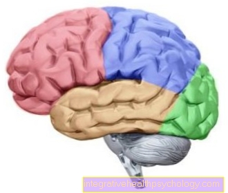
Brain lobe
Frontal lobe = red (frontal lobe, frontal lobe)
Parietal lobe = blue (parietal lobe, parietal lobe)
Occipital lobe = green (occiptital lobe, occipital lobe)
Temporal lobe = yellow (temporal lobe, temple lobe).
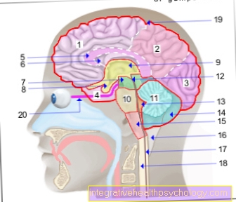
Cerebrum (1st - 6th) = endbrain -
Telencephalon (Cerembrum)
- Frontal lobe - Frontal lobe
- Parietal lobe - Parietal lobe
- Occipital lobe -
Occipital lobe - Temporal lobe -
Temporal lobe - Bar - Corpus callosum
- Lateral ventricle -
Lateral ventricle - Midbrain - Mesencephalon
Diencephalon (8th and 9th) -
Diencephalon - Pituitary gland - Hypophysis
- Third ventricle -
Ventriculus tertius - Bridge - Pons
- Cerebellum - Cerebellum
- Midbrain aquifer -
Aqueductus mesencephali - Fourth ventricle - Ventriculus quartus
- Cerebellar hemisphere - Hemispherium cerebelli
- Elongated Mark -
Myelencephalon (Medulla oblongata) - Big cistern -
Cisterna cerebellomedullaris posterior - Central canal (of the spinal cord) -
Central canal - Spinal cord - Medulla spinalis
- External cerebral water space -
Subarachnoid space
(leptomeningeum) - Optic nerve - Optic nerve
Forebrain (Prosencephalon)
= Cerebrum + diencephalon
(1.-6. + 8.-9.)
Hindbrain (Metencephalon)
= Bridge + cerebellum (10th + 11th)
Hindbrain (Rhombencephalon)
= Bridge + cerebellum + elongated medulla
(10. + 11. + 15)
Brain stem (Truncus encephali)
= Midbrain + bridge + elongated medulla
(7. + 10. + 15.)
You can find an overview of all Dr-Gumpert images at: medical illustrations
Prefrontal cortex
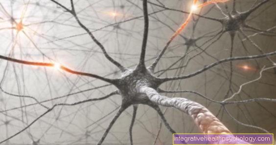
The turns of those parts of the frontal lobe that lie far forward are summarized as the Prefrontal cortex together. At these points you will find i.a. active thought processes e.g. a tricky math problem: the contents of the short-term memory take precedence over the intellectual eye examined. The information flies through the interaction of several nerve cells (Neurons), which form neuron loops like at the roundabout on the street, criss-crossing the cortex (cerebral cortex)! The mental contents are coded in the form of the electrical excitation of the neurons.
In addition to this, the prefrontal cortex probably plays a role as part of the Limbic system (see below, assignment but controversial), plus it contains the incorporated (internalized) values and social norms of one's own society. After all, you need those parts that are directly above the eye socket (orbit) (orbital prefrontal cortex) as a high-ranking member of the motivational circuit (reward system).
Olfactory cortex
At the base of the frontal lobe there are also phylogenetically old components (olfactory cortex, paleocortex and archicortex) that are dedicated to the sense of smell (olfactory sense) (see also olfactory tract). Presumably, the olfactory sensations in the so-called "primary olfactory cortex" (prepiriform cortex, also located to a small extent next to the frontal lobe in the temporal lobe) become conscious, the further assignments, a comparison with known sensations etc. take place in the adjacent "secondary olfactory cortex".
Note processing of sensory perception
Incidentally, this represents a widespread principle in the brain: all sensory perceptions come into consciousness at their primary cortical fields, but the integrative / analytical interpretation takes place in secondary fields and downstream associative fields. This thought is important because both types of cortex can have disorders independently of one another (see below agnosia, neglect). At least secondary fields are usually in direct proximity to the primary fields!
Incidentally, secondary olfactory cortex areas overlap on the orbital prefrontal cortex with the secondary centers of the sense of taste (see island cortex below). In general, these two senses are close to each other ("lower senses") and are afflicted with emotions and great willingness to act through the limbic system (see below) and the motivational circuit.
Example smelling
Everyone has this experience in everyday life: wherever it's good smells, you run there faster as if by yourself!
Basal forebrain structures
They are also located at the base of the frontal lobe, but in the form of core areas and not in the cortex basal forebrain structures. A core area of them, the nucleus basalis (nucleus Meynert) is to be understood as the link of the limbic system (see below) with several parts of the cerebral cortex. In this way, complex behaviors are influenced; it should also be important for learning (see below. Alzheimer's disease).
The is also particularly important on the frontal lobe Precentral gyrus (Motocortex, primary somatomotor cortex), because it serves as the uppermost center of any consciously planned movement (voluntary motor skills). It is surrounded by "premotor" and further "supplementary motor" cortical fields towards the forehead, which have a regulatory function in interaction with the pons (brain bridge) and cerebellum or prepare movements in an organizing manner. The frontal eye field (frontal center of vision) joins the forehead again. Here, arbitrarily targeted eye movements (saccades) are generated. The precentral gyrus is formed by the noticeable central sulcus Postcentral gyrus (primary somatosensitive cortex) separately. The latter is the important provisional terminus of most of the human sensations such as pain (protopathic sensitivity), tactile sensation (exteroception), sensation of position of the musculoskeletal system (Proprioception) and some others. It is only at this point that the aforementioned sense qualities come into our consciousness, even if initially without interpretation. Incidentally, the transverse central sulcus separates the motor cortex from the primary somatosensitive cortex and also separates the frontal lobe from the parietal lobe!
Another prominent furrow that Lateral sulcus, separates the lower parts of the frontal and parietal lobes from the temporal lobe. If one were to push a finger into the temporal sulcus, the lower surface of the finger (palmar surface) would brush over certain turns that belong to the temporal lobe. They are in different spatial orientation to the other turns of the temporal lobe and were therefore called "Gyri temporales transversi" (Heschl cross turns) designated.
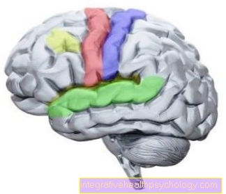
Important brain centers
Red = Gyrus precentralis, center for motor skills (movement)
Blue = postcentral gyrus, center for sensor technology (feeling / sensory perception)
Green = Wernicke - language center, center for language understanding
Yellow = Broca - language center, center for language articulation
Auditory cortex
These convolutions represent nothing less than the primary auditory cortex (auditory cortex), the temporary end point of a long auditory pathway that begins in the inner ear with the connection of the 8th cranial nerve (cochlear nerve) to the sensory cells (hair cells in the organ of Corti). Similar to other sensory qualities, the primary perception of tones, sounds, noises etc. has absolutely nothing to do with interpretation, i.e. an understanding evaluation and assignment. Words, melodies and the like can therefore only be interpreted in the interaction of the primary auditory cortex with so-called secondary cortical fields, in this case the secondary auditory cortex. Fortunately, this is located directly outside (laterally) adjacent to the primary auditory cortex! Our two secondary auditory cortexes (one per hemisphere of the brain) have the special feature that they have different focuses in relation to the processing of acoustic stimuli.
Note Dominant hemisphere
Rational language content such as a discussion about mathematics tends to be processed on the dominant hemisphere, artistic content such as music on the non-dominant side. By definition, the half of the brain (hemisphere) that mainly processes language is called dominant. With right-handers this is usually the left hemisphere and with left-handers it is variable with a slight numerical overhand also left.
After all, the secondary auditory cortex on the dominant side is called the “Wernicke language center”; this is where the understanding of the language takes place. The secondary acoustic cortical fields can be found directly outside on the lateral sulcus in the temporal lobe, more precisely in its uppermost turn (gyrus temporalis superior).
While the understanding of the language takes place here (the sensory component of speaking), the design of wording and sentence structure (motor component of speaking) takes place in parts of the lowest turn of the frontal lobe (gyrus frontalis inferior), the Broca language center. Failures at the Broca Center and the Wernicke Center result in different types of speech disorders (aphasia, see below).
Below the superior temporal gyrus lies the superior temporal sulcus of the same name. This furrow extends to the parietal lobe and is wrapped in a C-shape by one of its turns, the angular gyrus. The angular gyrus is an important interface between the secondary visual cortex (see below) and the secondary auditory cortex. In it, what is seen is provided with linguistic terms, corresponding disorders (alexia, agraphia and the inability to name the banal things that one sees, see below) are typical.
Another well-known area in the parietal lobe joins the postcentral gyrus posteriorly (caudally).
Read more on the subject here: Long-term memory
Note orientation in the brain
In the cerebrum and the diencephalon, terms such as “caudal = otherwise below”, “ventral = otherwise in front”, “dorsal = otherwise behind”, “oral / rostral / cranial = otherwise above” mean something different than in the rest of the body. This is because, during development, the cerebrum and diencephalon bend forward and the brain stem = midbrain + pons + medulla oblongata remain in the vertical direction of the spinal cord.
The usual axis is called the Meynert axis, the exception on the cerebrum and diencephalon is called the Forel axis. In relation to the latter means "caudal = behind", "ventral = below", "dorsal = above" and oral / rostral = front).
This area is called the posterior parietal cortex and is essential for orientation in three-dimensional space (spatial disorientation after a defect).
Caudal to the Broca center, directly above the temporal sulcus, the secondary somatosensitive cortex is part of the parietal lobe. Here the sensations that were listed above for the primary somatosensitive cortex are assigned to our wealth of experience and recognized (in the case of damage, “tactile agnosia, neglect, see below).
Visual cortex
In the occipital lobe, the extremely complex sense of sight (visual sense) is represented cortically. The visual pathway begins with the sensory cells of the retina and runs as the second cranial nerve (optic nerve) via a few intermediate stations to the primary visual cortex (visual cortex). In the simple representation of the brain from the side, this represents the most caudal (here: rear) pole (occipital pole) of the brain. Only a longitudinal section (median section) through the brain makes its entire extent clear, it runs in the wall of the sulcus calcarinus up to the border of the occipital lobe on the cingulate gyrus (represents a separate lobe, see below). Posteriorly (here: above), in the median section, the paietooccipital sulcus separates the occipital lobe from the parietal lobe. Both of the aforementioned furrows delimit a wedge-shaped section of the occipital lobe, the cuneus! In addition to parts of the primary visual cortex, this also contains the secondary visual cortex and other visual cortex fields, e.g. Generate eye-tracking movements (optokinetic reflex).
Repetition of the visual cortex
To repeat: what is seen becomes conscious in the primary visual cortex, Interpretation and analysis (e.g. to recognize writing) in the secondary visual cortex. In order to understand what is visually recognized, a fiber connection is absolutely necessary secondary visual cortex with the Wernicke Center (secondary auditory cortex).
In this connection, the angular gyrus represents an indispensable intermediate station. Understanding, however, is not to be equated with the ability to name in order to express what is seen in words, there must urgently be a connection from the Wernicke center to the Broca center, from there the premotoric center and to control motorized bark fields. At the end there is the activation of the corresponding Musculaturethat allows language formation (phonation and articulation).
Island bark
We spoke of the temporal sulcus earlier in the text. If you slide your finger far enough into this furrow, your fingertip hits it Island bark (own lobe, insular lobe). It is a field of bark that is dedicated to several sensory qualities (multi-sensory cortex), the sense of taste (gustatory sense), the Sense of balance (vestibular sense) and the very special sensitivity of the viscera (visceral sensitivity). Thus it represents the preliminary end point of the taste path, the primary gustatory cortex (becoming conscious). In addition, part of the primary vestibular cortex (becoming conscious) is located here. Likewise, especially on this bark of feelings like a filled one bladder, Nausea or a feeling of fullness after a long meal. It is information about the state of our internal organs, more primary visceral-sensitive cortex. As with other sensory qualities, the associated information runs through a well-defined path through the body (viscero-sensitive path).
Limbic system
A knife is inserted into the interhemispheric fissure (fissura longitudinalis cerebri) and cuts in the direction of the Brain stem (Median section), you can see numerous structures that can be attributed to the Limbic system (Limbik). It deals with emotions as well as with instinctual and intellectual behavior. Rather primitive services such as affective behavior in the context of self-preservation / species preservation and memory functions for different memory contents are thus decisively processed here. In addition, internal bodily functions (vegetative functions) are controlled here, certainly closely based on our emotions.
Note Limbic system
From such connections it is explained that e.g. the feeling of anger and anger can "hit you on the stomach"!
The following structures are included in limbic: Hippocampus (with gyrus dentatus and fornix), gyrus cinguli (own lobe of the cerebrum), gyrus parahippocampalis with area entorhinalis, corpus amygdaloideum (amygdala). Corpus mammilare (belongs to the diencephalon).
For functional reasons, it also includes parts of the olfactory brain, the indusium griseum, parts of the thalamus (belongs to the diencephalon) and the prefrontal cortex (see above). The limbic system owes its name to the spatial arrangement in the brain, because it swings like a fringe around the bar (corpus callosum) and the diencephalon. The bar is the largest fiber connection (i.e. white matter) between the left and right hemispheres of the cerebrum (comissure fibers) and synchronizes these with each other like a large bridge between two different cities. If it is cut, complicated symptoms occur, which the division of our cerebrum into two illustrates in an astonishing way (splitbrain). In any case, the cingulate gyrus lies on the bar (dorsal), parts of the diencephalon are embraced by the hippocampus with fornix, as far as the positional relationship! Parts of the limbic just mentioned are also vital in connection with the extensive memory that we have. Our Short term memory can store little information for seconds to minutes and is mostly located in the prefrontal cortex, but also in parts of the entire cerebrum. Now it often happens, however, that we want to memorize the information we are currently dealing with for a longer period of time, that is, we want to "learn" (consolidate memory). For this Learn is the Hippocampus and certain nerve connections (Papez neuron circuit and certain deviations from it), which contain large parts of the limbic, are indispensable. Damage in these areas causes the loss of memory or accessibility of information and other forms of "amnesia". A functioning hippocampus with a downstream limb transfers the information from short-term memory to long-term memory, where it can linger for several decades. The Long-term memory corresponds to a performance of the cerebrum as a whole and, for special matters, additional centers. When we spoke of information, we only meant factual information (explicit memory content) such as facts and events. The mechanisms for motor learning, the learning of courses of action as well as habits or even emotional learning (all implicit memory contents) also require the help of other special brain centers, which will not be discussed at this point.
Basal ganglia
Finally, we do not cut the cerebrum with the length of the interhemispheric gap, but rather in the middle across it, parallel to the forehead (frontal cut). With this incision, too, it is noticeable that some gray matter is embedded in the white matter of the cerebrum, which is not part of the cortex. The ancient anatomists called some of these nuclei "Basal ganglia“And over time this term has been expanded for functional reasons. Today one usually counts: the striatum with nucleus (Ncl.) Caudatus and putamen, the pallidum, the Ncl. subthalamicus and the substantia nigra. The striatum and pallidum lie to the side of the thalamus of the diencephalon, the Ncl. The subthalamicus is (as the name suggests) below the thalamus, while the substantia nigra is located far away in the midbrain. The exact interconnections of these areas and their integration into the rest of the brain fills entire textbooks; we reduce here to a practical level. As a whole, the basal ganglia control the extent, force, direction and speed of a movement that is still in the planning stage. What is special, however, is that they simultaneously evaluate the action, i.e. whether it could be useful in the overall context or not, or whether it is socially acceptable at all. One could say that they are also an extension of one's own values, which can curb inappropriate behavior.
Note basal ganglia
If you commit a shameful act, one or the other will probably notice a certain inner hesitation beforehand. Or the other way round: The starving person is particularly ready to drive because the basal ganglia “notice” this malady and encourage all actions.
Given these considerations, it is not surprising that some parts of the basal ganglia are important members of the motivational circuit. As such, they are constantly informed about possible upcoming rewards or displeasure in the absence of rewards, which they take into account in their processing of a movement. Especially when it comes to the topic Addiction as an extreme form of reward, they play a major role. In planning a movement, the basal ganglia are one of the three main routes of information flow, which begins with the will of voluntary movement in the limb. Typical diseases that are associated with a disorder of the basal ganglia are the Parkinson's disease and choreic disorders like Chorea huntington.
Common illnesses
Neurodegenerative diseases such as Parkinson's disease, Huntington's chorea, Alzheimer's disease and strokes, a headache, Epilepsy and Brain tumors occur comparatively frequently. With an increasing tendency one can find in our modern society depressions, Psychoses like that schizophrenia as well as addictions.
Other diseases or consequences of diseases of the cerebrum are:
- Multiple sclerosis (MS)
- Amyothrophic lateral sclerosis (ALS)
- Spasticity
- paralysis
- Eye paralysis
- Paresis
- Facial palsy
- Hemiparesis
- Hydrocephalus (water head)
- Encephalitis
- Prion diseases
- concussion
- Intracranial bleeding (ICB = Cerebral hemorrhage)
- traumatic brain injury
- Visual field loss
- Neglect
- Agnosia
- Alexia
- Agraphy
- aphasia
- amnesia