Heart sounds
General

Heart sounds are present in every healthy person and arise during the action of the heart. During the physical examination with the stethoscope, the Auscultation, thus, among other things, possible damage to the heart valves and cardiac arrhythmias can be detected.
A total of two heartbeats, and possibly up to four in children and adolescents, can be heard. The so-called heart murmurs are to be distinguished from this. They always have disease value and can, for example, provide evidence of a heart valve anomaly.
sdj
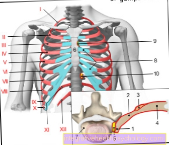
Heart anatomy
Our heart is one hollow muscular organ and can be turned into a left and right half organize. Both left and right halves of the heart have two so-called Cardiac cavities, the Forecourt (lat. Atrium) and the chamber (lat. Ventricle). So overall it results four heart cavities.
We continue to find four heart valves, which are essential for the orderly blood flow within the heart and in the vessels near the heart are. Thus chambers and atria are each separated by a so-called sail flap Cut:
- Mitral valve between the left ventricle and the left atrium
- Tricuspid valve between right chamber and right atrium
Because of her characteristic appearance, are the valves between the heart chamber and the large outflow channels Pocket flaps called:
- Aortic valve between left chamber and aorta (" artery")
- Pulmonary valve between the right ventricle and the pulmonary trunk ("pulmonary artery")
You can find out more about the anatomy of the heart here.
Illustration heart

- Right atrial -
Atrium dextrum - Right ventricle -
Ventriculus dexter - Left atrium -
Atrium sinistrum - Left ventricle -
Ventriculus sinister - Aortic arch - Arcus aortae
- Superior vena cava -
Superior vena cava - Lower vena cava -
Inferior vena cava - Pulmonary artery trunk -
Pulmonary trunk - Left pulmonary veins -
Venae pulmonales sinastrae - Right pulmonary veins -
Venae pulmonales dextrae - Mitral valve - Valva mitralis
- Tricuspid valve -
Tricuspid valva - Chamber partition -
Interventricular septum - Aortic valve - Valva aortae
- Papillary muscle -
Papillary muscle
You can find an overview of all Dr-Gumpert images at: medical illustrations
Cardiac function
To the Origin of the heart sounds to understand, it's worth a quick look at that How our heart works to throw.
Put simply, the organ can be called muscular pump consider: the left heart is pumping oxygenated blood in the big Body circulation, the right heart pumps again deoxygenated blood in the small Pulmonary circulation. Both parts are functionally linked in such a way that the carries the same amount of blood becomes.
Oxygenated blood flows from the superior and inferior vena cava (lat. Superior vena cava and inferior) in the right atrium. Through the opened tricuspid valve, the blood enters the right ventricle. From there it flows through the open pulmonary valve into the pulmonary arteries.
That in the lungs oxygenated blood gets through the pulmonary veins into the left atrium and then flows through the opened tricuspid valve into the left ventricle. Through the opened aortic valve, the blood gets into the artery (lat. aorta) and from there into the whole body directed.
During the Tension and expulsion phase, the blood flows from the chambers into the vessels near the heart.In order to build up the necessary pressure, there is a Tension of the heart muscle. This phase is known as Systole. As diastole on the other hand, is called the Relaxation and filling phase. Both chambers of the heart then fill with blood from the atria.
1. Heart sound
ly the first heart sound through the End of the sail flaps (Mitral and tricuspid valves). Furthermore, a Tension of the heart muscles watch while closing the flaps at the same time. This is how it turns out Heart wall in vibration and the first heartbeat becomes audible. So sometimes he will "Tension tone" called.
The best thing to do is let himself be Area of the apex of the heart or that fourth, left intercostal space wiretap. When the first heart sound particularly loud is, can be Narrowing of the mitral valve opening present (mitral valve stenosis). Furthermore, a to fast heart rate (lat. Tachycardia) Cause of a very loud first heart sound be. At a to the faint first heartbeat, can a Mitra valve weakness (Mitral regurgitation).
2. Heart sound
To Beginning of the relaxation phase, close the Pocket flaps (Aortic and pulmonary valves). This gets the Walls of the aorta and pulmonary artery in vibration and generate the second heart sound. In order to hear the second heart sound well, the doctor puts the stethoscope into the right, second intercostal space next to the Sternum.
Usually, that includes Aortic valve in front of the pulmonary valve. At our inhalation, this effect is amplified, making it a "Splitting" of the second heart sound can come.
A constant, breath-independent cleavage, however, is always morbid and can note for congenital Heart defect or Heart failure be. A louder, second heart sound speaks for arterial hypertensionwhile a quieter, second heart sound for one Narrowing of the opening of the aortic valve (s. Aortic stenosis).
Extra tones
Flap opening tone
When the miter valve stops in its opening movement, a flap opening sound can occur. These incomplete opening is usually through a Mitral valve stenosis caused.
3. Heart sound
Healthy adults usually do not have a third heart sound. At Children and adolescents however, it can be audible and is a consequence of the passive blood influx into the heart chamber. In adults, a third heart sound can especially occur Indication of heart failure ("Weak heart").
4. Heart sound
The fourth heart sound is also not in healthy adults audible. If it does occur, e.g. Heart failure or high blood pressure be. In some cases, he goes in healthy children and adolescents ahead of the first heartbeat.


.jpg)