histology
synonym
Microscopic anatomy
English: histology

Definition - what is histology anyway?
The word is histology consisting of the Word "histos", what in Greek "tissue" means and that Latin word "logos" for "teaching". So in histology "Tissue theory", one uses in everyday life technical aids such as a light microscope to use the Recognize the structure of different structures. Furthermore, in microscopic anatomy, organs are broken down into smaller and smaller components:
- histology - The tissue theory is an important part of medicine and biology, or anatomy or pathology.
- cytology - Cell theory deals with the functional composition of cells.
- Molecular biologye - The cells, in turn, are made up of many small molecules (particles).
Why do you need histology in everyday medical / biological life?
Your tasks are the Early diagnosis of ridges (Tumors) and whether this benign or malignant are, as well as the Detection of metabolic diseases, bacterial, inflammatory or parasitic diseases. In addition, the tissue theory contributes to Therapy decision at and has many other tasks in the clinic and in everyday research.
How does it all work now?
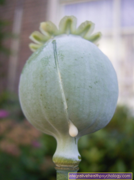
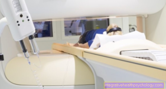
Of the Pathologist receives a Tissue sample, for example by trial excision stomach, Intestines, liver etc., or a piece of "tumor" that was surgically removed from an organ and makes micrometer-thin cuts. These will colored and can be examined with the light microscope or additionally with the electron microscope. The latter is very high-resolution and is mainly used in research.
Histotechnology
Histotechnology deals with the exact Processing of the fabricbefore it can even be examined. This is usually done by the medical technical assistant in the laboratory (MTA) responsible. These include: the fixation of the Tissuewhich is used for stabilization; the macroscopic (performed by eye) Looking at the fabric, as well as its Cuttingwhich of a Doctor running becomes; The Drainage and impregnation of the fabric in liquid paraffin; the Block the tissue sample in paraffin; the Cut out 2 - 5 µm thick sections as well as that Attach to the glass slide and finally that Coloring the cuts.
The Routine method in histotechnology is that Manufacture of an FFBE preparation, i.e. a paraffin-embedded tissue fixed in formalin, which is then stained in hematoxylin-eosin. This process lasts approximately from the sampling to the result of the analysis (diagnosis) a day or two.
Rapid section examination
This is required if the Surgeon needs information about the removed tissue during an operationto decide the course of the procedure. For example, a small, malignant tumor emerges from the kidney operated out. The quick cut is now required to see whether the tumor has already been completely removed or whether there is still a lot of malignant tissue on the edge zones of the tissue samples. In the end, the outcome of the high-speed section examination determines the course of the operation and the patient's further therapy plan.
How does a rapid section examination work? Within 10 mins becomes at -20 ° C the tissue is stabilized by freezing, then on so-called Microtome a 5 - 10 µm thick section is made. This is on a microscope slide, a small glass plate and colored quickly. At the end there is still the Diagnosis under the microscope and the The result can be forwarded to the operating room immediately.
Staining methods
Many histological staining methods have evolved over the past 120 years. The cell structures and tissues are divided based on the color reaction with the dyes basophilic, acidophilic, and neutrophilic cells. Furthermore still exist agyrophilic and nucleophilic structures.
Basophil colors all that one Acid group includes and with a basic dye (for example hematoxylin or methylene blue) is colored. Of the Cell nucleus is, among other things, basophil.
Structures that acidophilic are, are basic and can therefore be treated with eosion or acid fuchsin (acidic dyes) stain. This includes that Cytoplasm as well as collagen fibers. Neutrophils or lipophilic components cannot react with either an acidic or a basic dye and so can themselves do not stain.
Agyrophilic components can bind silver ions and convert it to elemental silver. A nucleophilic (nucleus = cell nucleus, nucleus-loving) color reaction is created by nucleophilic dyes in the cell nucleus. These are DNA-binding or basic substances that bind to nucleic acids.
The tried and true chemical dyeing methods are today complemented by immunological methods. The Antigen-antibody reaction this technology is used to prove certain cell properties. The reaction can then be made visible through a sophisticated technique.
Frequently used staining methods are:
HE staining = hematoxylin-eosin staining: Hematoxylin, a natural dye, colors all structures blue that are basophilic (= basic loving) and therefore acidic, such as the DNA, cell nuclei, ribosomes, etc.. Eosin, on the other hand, is produced synthetically. Eosin colors all cell structures red if they are acidophilic (= acid-loving) or basic. The Proteins of the cytoplasm, mitochondria, and collagen are among them.
Azan stain: It is made up of the first letters of both colors, azo carmine G and aniline blue-gold orange: This allows the Color the nucleus and muscle fibers red and the cytoplasm reddish. Collagen and reticular fibers will be at this staining blue.
The Giemsa stain (Giemsa's azure-eosin-methylene blue) is used Staining of blood cell smears. Cell nuclei can be easily recognized by the purple-red color reaction. The cytoplasm colors itself bluish a.
In the Elastica staining (Resorcinol fuchsin / orcein) all elastic fibers are shown in black and purple.
The van Gieson staining method is characterized by the fact that hematoxylin is used first. Then picric acid fuchsin (picrofuchsin) or picric acid thiazine is used. This is how they end up Cell nuclei black-dark brown dar, that Cytoplasm looks more like light brown. The counterstaining with picric acid fuchsin colors the elastic fibers and the muscle tissue orange-yellow and the collagen fibers red.
In the Trichrome staining according to Masson-Goldner the dye molecule size is the most important factor in the staining method. Iron hematoxylin is used, usually with three additional coloring agents, namely acid fuchsin, orange G and light green. It is colored collagenous connective tissue and mucus green on, Cell nuclei blue-black, Cytoplasm red, Muscles pale red and Red blood cells (Erythrocytes) Orange red on.
There is also one Grief stainingthat to Differentiation of bacteria serves. Gram-positive bacteria are colored blue and Gram-negative bacteria are colored red.
The Ziehl-Neelsen staining will also be at bacteria namely with those who are acid-proof, used and provides For example, tuberculosis pathogens are shown in red.
Other colors that should be mentioned here are the Berlin blue Reaction which for the Detection of trivalent iron ions in the tissue section is responsible and the Iron hematoxylin staining method according to Heidenhain.