Knee pain on the inside
introduction
Under a internal knee joint pain refers to a pain that is mainly (not always exclusively) concentrated on the inner part of the knee joint.
This includes pain in the area of the inner thigh and lower leg, the Inner band, the surrounding soft tissues and the internal knee joint space.
Knee joint pain on the outside can be caused by direct damage to the anatomical structures involved or as transmitted pain in the event of damage at an anatomically distant location.

Please note
In no case does the “self” diagnosis replace a visit to your trusted doctor! We also make no claim to the completeness of the differential diagnoses presented (alternative causes). We assume no liability for the correctness of the self-diagnosis you have made! We strictly reject any form of self-therapy without consulting your doctor!
Anatomy of the knee joint

- Thigh bone (femur)
- Inner meniscus
- Outer meniscus (outside)
- Fibula
- Shinbone (tibia)

I would be happy to advise you!
Who am I?
My name is I am a specialist in orthopedics and the founder of .
Various television programs and print media report regularly about my work. On HR television you can see me every 6 weeks live on "Hallo Hessen".
But now enough is indicated ;-)
The knee joint is one of the joints with the greatest stress.
Therefore, the treatment of the knee joint (e.g. meniscus tear, cartilage damage, cruciate ligament damage, runner's knee, etc.) requires a lot of experience.
I treat a wide variety of knee diseases in a conservative way.
The aim of any treatment is treatment without surgery.
Which therapy achieves the best results in the long term can only be determined after looking at all of the information (Examination, X-ray, ultrasound, MRI, etc.) be assessed.
You can find me in:
- - your orthopedic surgeon
14
Directly to the online appointment arrangement
Unfortunately, it is currently only possible to make an appointment with private health insurers. I hope for your understanding!
Further information about myself can be found at
To the diagnostic
Using our “Self” diagnostic agent is simple. Follow the respective link offered, where the location and description of the symptoms best match your symptoms. Pay attention to the point of the knee joint where the pain is greatest.
Where is your pain?
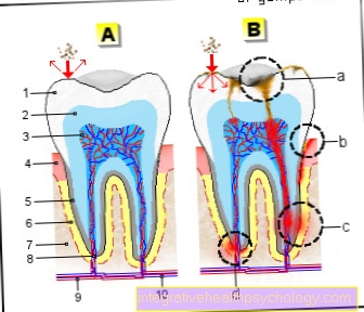
Illustration internal knee pain

A - Right knee joint from the front
- Inner thigh muscle -
Vastus medialis muscle - Femur -
Femur - Kneecap -
patella - Inner meniscus -
Meniscus medialis - Inner band -
Ligamentum collaterale tibiale - Internal calf muscle
Gastrocnemius muscle,
Caput mediale - Shin -
Tibia
a - Inner ligament injury
(Inner ligament tear)
b - tear of the medial meniscus
c - medial
(internal) knee osteoarthritis
You can find an overview of all Dr-Gumpert images at: medical illustrations
Medial meniscus tear

- Synonyms:
External meniscus rupture, external meniscus lesion, external meniscus degeneration, external meniscus damage, internal meniscus disease - Place of greatest pain:
In the area of the inner knee joint gap. - Pathology / cause:
Accidental or wear-related (degenerative) tear of the inner meniscus. - Age:
every age - Gender:
no gender preference - Accident:
Twisting trauma (accident) of the knee joint during exercise. With degenerative cracks, there is usually no memory trauma. In severe injuries, common occurrence with torn inner ligament and anterior cruciate ligament tear (unhappy triad) - Type of pain:
Stinging, light to dull, drawing. Possibly. limited, partially blocked mobility of the knee joints. Incoming stabbing pain after turning the leg. - Pain development:
After a trauma (accident) suddenly, otherwise slowly increasing or recurring with breaks. - Pain occurrence:
V.a. during exercise, in a crouching position or after unfavorable rotational movements of the knee joint. If the meniscus becomes trapped in the knee joint, the knee can no longer be fully extended and constant pain can result. - External aspects:
In the case of acute injury, swelling usually increases. In the case of degenerative cracks, there is less swelling depending on the load, sometimes none. - Further information:
You can find comprehensive information under our topic: Meniscal tear
Medial (internal) knee osteoarthritis

- Synonyms:
Arthrosis of the inner compartment, medial gonarthrosis - Place of greatest pain:
In the area of the internal knee joint gap. - Pathology / cause:
Wear-related (abrasion) cartilage damage with inflammation of the mucous membrane with main damage in the area of the inner knee joint. - Age:
Older age (> 50 years) - Gender:
no gender preference - Accident:
No. However, "activation" of an existing, dormant osteoarthritis is possible through a knee injury. Possibly. Condition after a previous partial removal of the meniscus. - Type of pain:
Stinging, light to dull, drawing. Stiffness of the knee joint. Possibly. limited mobility of the knee. - Pain development:
Slowly increasing, sometimes stabbing, sometimes pulling depending on the stage of osteoarthritis. - Pain occurrence:
Morning start-up pain. Increase in pain under exertion (with increasing walking distance). - External aspects:
Swelling, possible overheating. Often bow legs (genu varum). - Further information:
You can find comprehensive information under our topic: Osteoarthritis of the knee
Inner ligament injury

- Synonyms:
Internal ligament tear, internal ligament injury, damage to the medial collateral ligament - Place of greatest pain:
In the course or at the beginning / origin of the inner band - Pathology / cause:
Overstretching or tearing of the outer ligament.
Further information on the topic: Inner ligament stretch in the knee - Age:
Mostly young, physically active people. - Gender:
no gender preference - Accident:
Yes. Usually it is a so-called varus trauma. This means that the knee joint is forcibly pushed into a knock-kneed position. If the stretch reserve of the outer ligament (ligamentum collaterale laterale) is exceeded, the ligament will tear or tear. - Type of pain:
piercing, bright. - Pain development:
Suddenly. Often in the context of football injuries. - Pain occurrence:
In connection with the injury. Pain when checking the inner ligament stability. Possibly. Inward instability of the knee joint. - External aspects:
Lateral, possibly general Knee swelling.
Mediopatellar plica

- Synonyms:
Shelf Syndrome - Place of greatest pain:
Inner knee joint portion, inner thigh roll. - Pathology / cause:
Mechanically irritating folds of the mucous membrane (lat .: Plica) of the inner knee joint, which can cause an inflammatory reaction and cartilage damage.
Internal knee pain after jogging If internal knee pain occurs after jogging, then this can be Beginners or after a long break be completely normal as long as this completely disappear overnight.
If this is not the case, the Pain a warning sign be that possibly a serious cause is behind it.
Keep the knee pain above you longer period then these should be clarified with an appropriate specialist.Knee pain can have a number of different causes. Perhaps the pain is one wrong running technique underlying. Your knees may be up when running bent too much, So that the Kneecap too much pressure is pressed into their bearings.
This kind of Running style is also called "sitting". It leads to excessive stress on the Knee joints.
Too intense training can also lead to knee pain after jogging. The pain is already kicking While the training on, you should get this abort. However, if the pain only occurs after training, you should use the Put your legs up and possibly the knee cool.
A Increase in training intensity can sometimes also cause inflammation of the Bursa to lead.
Also congenital deformities, like the well-known If one, can be a cause of internal knee pain. Most of the stress here is on the inner articular surfaceso that they are exposed to a significantly higher level of stress and strain than the outer joint surfaces. Due to the higher stress, it can lead to a increased cartilage abrasion what will ultimately come over time Knee osteoarthritis can lead.
Also a muscular imbalance the individual Muscles can lead to internal knee pain after jogging. Is the inner one Thigh muscles for example more pronounced than the external it comes to one Displacement of the kneecapso that it can no longer slip optimally through its sliding bearing and hit other joint surfaces.
Knee pain can also be caused by a instability in the Hip- or Ankle joint occur. If the muscles on the inside of the foot are too weak so that the foot bends inward, this affects the knee joint. However, it likely would more likely to cause pain on the outside of the knee joint to lead.Enter the internal knee pain repeated after jogging on, you should get one too Change of running shoes consider. They may be worn out or you can tell there are differences between the sides by the wear on the sole. Here it is recommended for one Treadmill analysis participate and possibly buy new running shoes. Another cause of internal knee pain can also be one Meniscus damage or a Damage to the tendons be considered. In the case of acute knee joint injuries, you can call yourself "Emergency response“To the so-called PECH rule hold: pause, ice, compression (against possible swelling) and elevation.
Osteoarthritis / meniscus damage
Of the meniscus represents a kind of disc-shaped cartilage in the Knee joint there is one Inside- and one External meniscus. They serve to compensate for the unequal joint shapes and serve the "Buffering“Of pressure loads on the joint surfaces.
Each meniscus is made up of three parts: a front horn, a rear horn and a middle part. In each of these proportions there can be Cracking come. Depending on the type of Crack shape For example, you can find basket handle, longitudinal or horizontal cracks. A meniscus damage can now lead to internal knee pain.
Damage can occur on the one hand external force / force arise or on the other hand by one long-term wear. Chronic overuse, for example by extreme exercise can lead to small, fine cracks, which over time may develop into one with normal movement larger crack or even one demolition being able to lead.
Meniscus damage often results from a Combination of turning and bending movements. The most common is then Medial meniscus affected, as this is fixed to the inner collateral ligament and is therefore particularly at risk of injuries. In the case of meniscus injuries due to rotational movements, there is often an additional one Injury to the anterior cruciate ligament. A classic injury involving the medial meniscus and the anterior cruciate ligament is the "Unhappy Triad Injury“In which the inner side ligament is also torn.The internal knee pain is mostly then stabbing or pulling. You can occur suddenly and very be strong and are often especially in Squatting position worse.
There may be one Restriction in knee movement, because parts of the meniscus block the movement. Stretching the leg in particular causes severe pain. Often there is also a so-called Relieving posture of the knee, keeping the knee joint flexed. In addition to the pain in the knee, restricted mobility and relieving posture, it can lead to a swelling come in the knee joint, which leads to a so-called "dancing patella" can lead.arthrosis represents progressive damage and ultimately the Breakdown of the articular cartilage Without the corresponding cartilage coating, the joint surfaces rub directly against each other, whereby the Pain caused in the joint. Especially on the back surface of the Kneecap and on the articular processes of the thigh bone it comes to Osteoarthritis im Knee joint.
There are basically two types that lead to joint arthrosis. Either osteoarthritis develops through the Consequences of an accident or through Cartilage wear in the course of life.
A Incorrect or overloading, for example caused by too intensive running training, but also Misalignments (here especially bow and knock knees) and eventual Previous damage in the knee joint area Tape- or Meniscal injuries, are very common Osteoarthritis trigger.
But also very overweight can through a high Cartilage wear lead to osteoarthritis of the knee. The internal knee pain in osteoarthritis is mainly in the area of the Joint space. They are often stabbing and significantly increase in intensity during exercise. In particular activities like Climb stairs are very painful for people with knee osteoarthritis, as the bending movement in particular causes pain in the knee.
If too much overload they can already at rest occur. Otherwise, osteoarthritis of the knee can often result in a so-called "morning start-up pain" determine. The osteoarthritis can persist through irritation ignite and to one arthritis lead, so that in addition to the pain there is also a swelling or joint effusion.Crack
A cracking noise when moving the knee can have a number of different causes. Possible Air pockets in the Synovial fluid, Cartilage damage, Damage to the ligaments, one Overloading the joint or one Knee osteoarthritis can be the cause of a cracking knee joint.
The most common is age-related wear and tear of the cartilage and bone make out such a crack in the knee.
The crunching or cracking noise is often an indication for Damage to the cartilage or signs of wear and tear. The constant bending and stretching movements lead to a heavy stress on the joint and the articular surfaces. In addition, the entire body weight rests on this joint. The cracking can also get through increased, excessive exercise caused. Athletes are therefore much more frequently affected by this than people who do little or no sport.
Past Knee injuries how Cruciate ligament tears or Injuries to the menisci can also lead to a cracking or snapping sound, often additionally accompanied by Knee pain are. The pain then mainly occurs Flexing movements of the knee joint.
Should the cracking after a Fall or a possible injury of the knee joint, it is advisable to consult a doctor directly. Under no circumstances should you wait to see whether the sound goes away again. One should also consult a doctor if the knee cracking is associated with knee pain and one swelling of the knee is.No matter what type of knee pain you are suffering from, there are still some basic principles you should keep in mind. It is always important to know what the knee pain is like. One differentiates whether the pain is more likely stabbing, pulling, oppressive, permanent or only from very short duration are.
The exact one is just as important Place of pain. A distinction is made between front, back and side knee pain. As an indication of the cause, the situation be when the pain occurs. A distinction is made between resting pain and pain that only occurs during exercise.treatment
If a prosthesis has to be used for treatment, rehabilitation should take place afterwards The treatment of internal knee pain can be conservative without surgery or minimally invasive Knee arthroscopy or major surgery. The choice of therapy depends on the root cause of pain Degree of damage, the corresponding Knee structure and the Wishes of the patient. For example, the pain may occur due to overuse after jogging. No acute treatment is required here, just taking it easy is enough to allow the internal knee pain to subside. It looks completely different with an inner meniscus tear.
Treatment of the medial meniscus
The treatment of the medial meniscus tear depends entirely on the age, mobility and physical activity of the patient. In the case of a slight tear, an attempt can first be made to treat the internal knee pain caused by it with the help of rest. In addition, the patient should always have a physical therapy in order to maintain the mobility of the knee joint even after the injury. It is also important that the patient does not put any strain on the knee if there is internal knee pain. Short-term walking on crutches is therefore a suitable treatment measure. If the medial meniscus tear causes a Joint inflammation the patient can treat internal knee pain with anti-inflammatory drugs called non-steroidal anti-inflammatory drugs (NSAIDs) and cooling. If the meniscus tear is too deep or too severe, it may be necessary to remove part of the meniscus. This treatment of internal knee pain caused by the meniscus tear should always be the last resort.
To the Medial meniscus to achieve a reflection (a so-called arthroscopy) of the knee joint. In addition to the kneecap, the surgeon places at least two small cutsthrough which a camera and tools can be inserted into the knee joint.
This minimally invasive approach enables not only the menisci but also the Cruciate ligaments or Mucosal folds are supplied. The advantage is that the patients recover much faster and there is a good cosmetic result. In addition, the rate of wound infections is significantly reduced compared to large open surgeries.
To the Complications is next to the failure above all that thrombosis to call. However, the risk is with less than 1% to be classified as low. It should be used for prevention Heparin- Abdominal injections are administered until the knee can be fully loaded again.If in the mirror one because of internal knee pain Tear of the medial meniscus is found, it will be taken care of immediately. It depends on the area in which the crack is located. Of the cartilage, which makes up the meniscus, is only well supplied with blood vessels at the base of the knee joint. This means that suturing the meniscus is successful in the case of tears close to the base.
After the operation, the knee joint must first be treated with a Stretching bar be kept quiet. For about 3 weeks Forearm crutches be used to avoid full knee strain. The diffraction is at the physiotherapy slowly rebuilt, but the patient should adjust to it up to dNo strenuous sports for three months to practice. Overall, the number of meniscus tears that can be treated with one suture is low, but the prognosis is very good.
For cracks further from the base, the blood supply is insufficient for the cartilage to heal. In this case a Partial meniscal resection performed. The torn part of the meniscus that causes the internal knee pain becomes away. Since the meniscus does not have to heal here, the knee can be loaded normally much earlier. Usually only small pieces of the meniscus are removed, otherwise an early one arthrosis of the knee joint.
Usually a knee examination is necessary. Treatment of Unhappy Triad
An injury that also involves the medial meniscus is the Unhappy triad. Next to the Medial meniscus and the inner sideband is that too anterior cruciate ligament affected. As described above, the medial meniscus is treated as part of a knee joint reflection. In addition, there is also replaced the anterior cruciate ligament. This is usually a piece tendon taken from the inside of the thigh and attached as a new cruciate ligament in the knee joint.
If the medial meniscus does not need to be treated, one waits first about 4 weeks until the swelling has subsided and examine the knee again. On the one hand, at this stage it can be decided whether the stability of the knee joint is sufficient for the patient's physical activity and therefore does not have to be operated on. On the other hand, delayed interventions on the cruciate ligament have a slightly better prognosis in terms of functionality.
The follow-up treatment includes gentle physiotherapy and the relief with crutches. After approx four months exercise can be started carefully.Treatment of torn ligament
Inner ligament tears as a cause of internal knee pain can in most cases conservative be treated. After the accident, the knee should be immobilized. Usually the doctor will prescribe one for this rail (also called an orthosis) that for six weeks will be carried. During this time, the inner band can sufficiently stabilize itself. Until the movement is completely restored physical therapy can three months pass away. If there is still a feeling of instability afterwards or the patient is very active in sports, the tendon can also be sutured through a small incision. But this is more likely to happen Combination injuries recommended as the Unhappy Triad.
Another way to treat internal knee pain is one minimally invasive surgery, in which the cruciate ligament is removed and usually replaced by a tendon from a muscle (for example the semitendinosus muscle). Since the new cruciate ligament cannot be fully loaded immediately, it takes a little longer for this treatment of internal knee pain to take effect.
Treatment of the mediopatellar plica
Since the Mediopatellar plica a fold of the mucous membrane without function for the knee, this can be done without any problems with a knee arthroscopy removed. The fold is directly at the level of the Joint capsule so that no more parts can be jammed when moving. Here, too, the pressure must then be relieved using forearm crutches until the knee can be relieved painlessly.
Treatment of osteoarthritis and cartilage damage
The arthrosis can with the exception of the Knee replacement not be cured. However, there are numerous ways to stop the course of the degenerative disease or at least to slow it down and thus eliminate the pain.
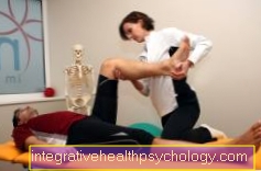
The first step of treatment is that conservative therapy. This includes in particular Movement without straining the knee joint. Especially Swimming and cycling are well suited sports to keep the joint moving. It is not advisable to do sports that involve rapid, abrupt changes of direction and heavy strain on the knee joint, such as Squash or soccer.
Additionally should Obesity be reduced, as the additional body weight further overloads the already damaged knee. In the physical therapy or knee training, which is often offered by health insurance companies, professionals teach patients exercises that they can do at home to further promote the mobility of their joints.In consultation with the doctor, the intake is also so-called Anti-inflammatory drugs advisable. These are pain relievers such as Ibuprofen or Diclofenacwhich, in addition to reducing the pain, also limit inflammation of the diseased joint. Using ointments with these ingredients or putting on wraps can also provide relief.
Especially if one Axial misalignment of the leg there is one Adjustment with insoles possible. With internal knee pain, the patient often has a If a. By getting the shoe sole increased on the outside, this can be partially offset.
The next step in therapy is the cold application in the form of a Cryotherapy or a heat therapy for application. In the early stages of osteoarthritis, heat has a pain-relieving effect. However, as soon as the osteoarthritis is activated, i.e. when inflammation has arisen due to the loss of cartilage, cold should be applied instead of heat.
An alternative to heat therapy is that Electrotherapy, whereby medium-frequency currents are applied to the surrounding tissue. The warming leads to increased blood flow and relaxation of the muscle tissue and, as a result, to a decrease in pain. Again, should no additional inflammation present in the arthritic joint.For some years now, the statutory health insurance has also been using the acupuncture reimbursed for knee osteoarthritis. Scientifically, a positive effect could be determined.
If no improvement in internal knee pain can be achieved with conservative measures, the joint will be mirrored during the further course of treatment Repair of the damaged articular cartilage into consideration. One possibility is that Microfracturing of the bone exposed through the cartilage atrophy. The doctor drills a few small holes in the bone as part of the reflection. These defects are replenished by the body. Since the bone is well supplied with blood, stem cells are also flushed out with the blood. From these a new replacement cartilage which, however, cannot keep up with the original cartilage in terms of resilience.
Another possibility is that Growing new cartilage cells. For this purpose, a piece of cartilage is removed during an initial mirroring, which is then reproduced in the laboratory within one to two months. Then it is brought back into the joint with a second reflection. With this procedure you can too major defects be covered.
A third alternative for more small cartilage defects is the Relocation of a cylinder of cartilage bone. A punch of cartilage and bone is taken from an area of the joint that is lightly stressed. The cylinder is then attached to the main load zone as a replacement.
All three procedures of cartilage replacement are mainly for younger patients well suited to delay the time until the joint replacement.At the same time, a Articulated toilet be performed. In this context, disturbing folds of the mucous membrane or bony attachments (Osteophytes), which can cause severe pain in addition to cartilage abrasion.
The final stage of osteoarthritis treatment is one surgery.
Especially in younger patients up to 60 years with a misalignment of the legs (in our case bow legs) is a Corrective osteotomy very promising. The bow legs result in one unilateral osteoarthritis on the inside of the knee joint, which also explains the increased internal knee pain.
With an osteotomy, that will Shin severed below the knee joint and then stretched until the leg axis is rather light X-leg shape is. The resulting gap is filled with bone from the iliac crest or artificial bone and then fixed with plates and screws. After the operation, the leg must six weeks relieved by crutches and mobilized with the help of the physiotherapists. During this time, the leg can slowly be fully loaded again under guidance. To about a year the metal parts are removed from the leg in a small operation.Another OP option is that Joint replacement. This is aimed for when the patient's pain cannot be remedied by other means and the quality of life is reduced due to the restriction of mobility.
A small form of joint replacement is that unicondylar sledge prosthesis. This prosthesis is characterized by its size. It only replaces the affected inner part of the knee joint, which is removed during the operation and with a Metal prosthesis and plastic is replaced. Another benefit is that faster rehabilitation after surgery. Requirements for a sled prosthesis is a stable ligaments, no misalignment of the leg and no excessive physical activity.
If these prerequisites do not apply or the rest of the knee joint already shows arthritic changes, this is one Total endoprosthesis (Knee replacement) the better choice.
With a total endoprosthesis, both the upper part of the knee joint, which is actually through the Thigh bone as well as replacing the lower part of the knee joint, which is the tibial head. The Bone parts are straightened and anchored the prosthetic parts with small wedges and bone cement.
After each form of prosthesis implantation, a Rehab carried out in order, on the one hand, to achieve complete mobility under professional guidance and, on the other hand, to be optimally prepared for everyday life. The prognosis for a knee replacement is good. she holds up to 20 years and enables painless full functionality of the knee joint.
Medical terms related to the knee joint
For the exact anatomical assignment, we refer to the Anatomy Lexicon our page.
Here are some key terms related to knee pain:
- Head fibulae - fibula head
- dorsal- back
- Fibula - fibula
- Femur - thighs
- Femoral condyle - thigh roll
- Gon - knee, knee joint,
- Hoffa - Fat body of the knee joint
- lateral - outside, on the side
- Medial collateral ligament - inner band
- Ligamentum collaterale laterale - outer band
- Ligamentum cruciatum anterius - anterior cruciate ligament
- Ligamentum cruciatum posterius - posterior cruciate ligament
- medial - Inside
- Medial meniscus - medial meniscus
- Lateral meniscus - External meniscus
- patella - kneecap
- Patellar tendon - kneecap tendon
- popliteal - concerning the hollow of the knee
- Quadriceps tendon - anterior femoral tendon
- Tibia - shin
- Tibial plateau - articular surface of the tibia
- Tibial tuberosity - Elevation of the kneecap tendon attachment to the shin
- ventral - front