Liposarcoma
definition
Liposarcoma is a malignant tumor of the Adipose tissue.
He kicks like everyone else Sarcomas, relatively rarely.
The fat cells do not develop in accordance with the norm, whereupon the degenerated cells become tumor arises.
Of the soft tissue sarcomas, liposarcoma is the second most common after malignant fibrous histiocytoma.
Liposarcomas are around 15% to 20% of soft tissue sarcomas. Rudolf Virchow was the first to describe liposarcoma as an independent disease in 1857.

Epidemiology / Occurrence
The tumor usually occurs in adults, but there have also been cases (about 60 in total) in which children and adolescents were affected.
The mean age of onset is 50 years. Liposarcomas most often occur between the ages of 50 and 70.
Men are slightly more likely to have liposarcoma than women. The incidence is around 2.5 new cases / 1,000,000 inhabitants / year.
causes
A clear explanation of the cause of the development of a liposarcoma is not yet known and is currently being researched.
Most tumors arise "de novo“By a degeneracy of embryonic progenitor cells.
However, there are theories about possible causes: There is a presumption that there is a connection with previous Radiation treatment with ionizing radiation.
In individual cases, a liposarcoma developed from a lipoma or from a burn scar. Whether the liposarcoma can have genetic causes is also not yet clearly confirmed, but it is suspected.
localization
Most often, liposarcomas develop deeply Soft tissue of the lower extremity (mostly on the thigh).
Second most often, they come in the upper extremity and the Retroperitoneum in front.
They can also occur on the trunk of the body.
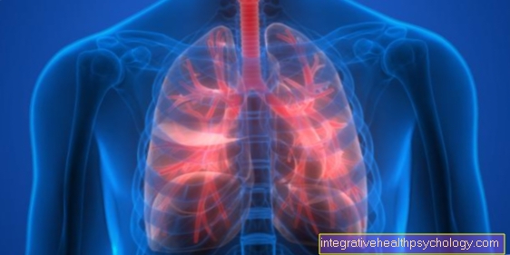
It occurs in 15% to 20% of cases Metastases which are most commonly found in the lungs. Metastases can also be found in the peritoneum, the diaphragm, the Pericardium, of the liver, the bones and the lymph nodes.
Occurrence on the thigh
Liposarcomas are particularly common (40%) on the thigh. Those affected notice a small bump or swelling under the skin. Usually, the thigh tumor does not cause pain.
Imaging tests such as CT, ultrasound, and MRI can provide clues as to whether it is a liposarcoma or a harmless fat tumor (lipoma). In addition, the actual size and extent of the tumor into the surrounding tissue are determined. For a more detailed examination, a tissue sample must be taken to determine the degree of malignancy.
Occurrence on the stomach
Liposarcomas can also form in the abdominal cavity or affect the gastrointestinal tract. Normally, the fatty growths here initially arise as a painless mass and it takes longer than elsewhere to be noticed and treated.
Liposarcomas that occur in the abdominal cavity can grow unnoticed over a long period of time and become very large without any external swelling. Often the patient only notices pain and see a doctor when the tumor has reached a considerable size and is pressing on surrounding organs or vessels in the abdomen.
The symptoms of liposarcoma in the abdomen are non-specific and depend on which organs are affected. The symptoms range from abdominal pain that cannot be precisely localized, digestive problems, constipation and nausea, to anemia.
If the liposarcoma cannot be surgically removed because it is very large and may have grown together with other organs or large vessels in the abdomen, the patient is given radiation therapy. The aim of this treatment is to reduce the size of the tumor so that it can then be completely removed by surgery.
pathology
Liposarcomas can be very large and heavy, depending on their location. Tumors of several kilograms Weight are not uncommon.
In extreme cases, they can weigh up to 30kg.
First a few words about the "macroscopic image“Of the tumor, that is, what the tumor looks like when seen with the naked eye.
Often times, the tumor appears well encapsulated and limited at first, but is occasionally in the vicinity of the main tumor no tumor deposits found.
Liposarcomas are yellowish in color (just like the fat tissue itself) and a gelatinous-mucous structure.
There are often in the tumor itself Necrosis (dead cell areas), Hemorrhages (Bleeding) and Calcifications.
The histological (microscopic) Image of the tumor describes what you see when you cut the removed tumor into fine layers and look at them under the microscope.
When looking at the so-called sectional images, several sub-types can be distinguished. These subtype classifications are also used to estimate a prognosis.
Here one indicates the degree of dedifferentiation. The more the cells are dedifferentiated, the greater the difference between the degenerate tumor cells and healthy cells and the worse the prognosis for the further course.
The "well differentiated“ (= little dedifferentiated) Liposarcoma is the most common at 40-45%.
The cells differ very little from mature healthy adipose tissue.
Dedifferentiation is moderately advanced. Synonyms for the "well differentiated“Liposarcoma are atypical or lipomatous tumor atypical lipoma.
The "Myxoids / round cell“Liposarcoma is the second most common at 30-35%. Dedifferentiation is already moderately to highly advanced.
The "Pleomorphs“Liposarcoma accounts for 5% of liposarcomas. Dedifferentiation of the cells is highly advanced.
The "Dedifferentiated“As the name suggests, liposarcoma is also highly dedifferentiated. But this only happens very rarely.
Symptoms
Liposarcomas often remain asymptomatic for a long time and therefore go unnoticed.
Depending on the location, the symptoms can vary.
Usually the first one is slowly growing solid tissue perceived. Depending on the depth of the liposarcoma, this increase in tissue will be noticed sooner or later.
If the tumor develops in the retroperitoneum, for example, it is usually diagnosed very late because it is hardly noticed there.
The main symptom for retroperitoneally located tumors are Abdominal discomfort (Abdominal discomfort), as the tumor begins to press on the organs as it grows.
The swelling of the limbs is usually noticed quite early.
Is the tumor adjacent to Nerve tracts it can press on them as it grows and thus become noticeable through tenderness.
are Blood vessels In the vicinity it can happen that these are compressed and it can cause discomfort in the blood flow in the affected area.
The larger the tumor becomes, the more likely it can lead to functional impairments (If you have a tumor in the thigh, for example, the leg can no longer be fully bent).
General symptoms, as they do with many Cancers occur can also be present in liposarcoma. These include, among others Weight loss, night sweats, fatigue, tiredness, nausea, and vomiting.
Pain as a symptom
Liposarcomas usually only cause pain when the tumor constricts organs or pinches nerves. Depending on the location and size of the tumor, it can exert pressure on various organs, which is noticeable through pain in the abdomen.
Pain can also occur when the liposarcoma compresses a nerve, which often causes tingling and numbness in the affected area of the skin.
Can a liposarcoma metastasize?
A liposarcoma can metastasize. In the process, small nests of tumor cells are detached, which can get into the bloodstream and thus spread throughout the body and form metastases.
Liposarcomas metastasize particularly frequently to the lungs, but bones, liver, peritoneum, diaphragm and the pericardium can also be affected.
Small metastases often cannot be detected by CT or MRI.
diagnosis
If an increase in tissue is noticed, imaging methods such as Computed Tomography (CT), the Magnetic resonance imaging (MRI), angiography or the Scintigraphy applied.
They should show how big the tumor is and how it relates to the surrounding structures (Vessels, nerves, organs) so that you can assess the possibilities for a removal. In addition, it is checked whether metastases have already formed in other regions.
If the diagnosis is to be secured, it is usually one biopsy with subsequent histopathological examination necessary.
Depending on the size of the tissue proliferation, only part of the knot or even the whole of it is removed.
After removal, this is cut into fine layers, which are then examined by an experienced pathologist under the microscope.
In addition to the histological examination, an immunohistochemical examination is carried out, which helps to distinguish the supposed liposarcoma from other sarcomas.
Different dyeing techniques are used. Express well-differentiated liposarcomas Vimentin and S-100. If only vimentin is expressed, this is a sign of a poorly differentiated tumor.
Diagnostics with ultrasound
Using an ultrasound from the abdomen, the doctor can assess whether a liposarcoma has formed there and whether metastases have developed. Metastases mostly affect the lungs, but can also occur on the liver, diaphragm, peritoneum or the pericardium.
Liposarcomas can be easily visualized and diagnosed with ultrasound, but more precise information about the malignancy can only be provided by a histological examination by the pathologist.
Diagnostics with MRI
Before taking a tissue sample (biopsy) for the histological examination of the tumor, an MRI should be done. Magnetic resonance tomography (MRT) is a high-resolution imaging method that is used to accurately assess the spread of the tumor.
It can also be determined whether blood vessels have already been infected. However, a definitive diagnosis can only be made by examining a biopsy.
therapy

If the location of the tumor allows, it makes most sense to have it surgically removed completely. This is also the best prevention against a relapse (= Recurrence) of the tumor.
A sufficient safety margin must be provided here so that the tumor cells are not carried over to other tissue during the operation and can continue to grow there.
If the removal is not possible because it has already infiltrated other areas (means has grown into this) or the dedifferentiation of the liposarcoma is too advanced, a radiotherapy be performed.
Even if the liposarcoma is considered to be the most radiation-sensitive sarcoma, scientific studies have not yet found any increase in survival time with radiation treatment. If metastases have already formed, there is a high probability that one will chemotherapy follow, but this is currently still in research.
forecast
In principle, liposarcoma is curable.
However, the chances of recovery depend on the size and condition of the cells (see pathology) of the tumor. Another important prognostic factor is whether metastases have already formed. At the "well differentiated“For liposarcoma, the prognosis is usually very good.
The 5-year survival rate is 88-100%. This means that after 5 years, 88-100% of the sick are still alive.
The good prognosis also comes from the fact that this form metastases rarely form. At the "Myxoids / round cell“Liposarcoma has a worse prognosis. The 5-year survival rate is only around 50%.
The "Pleomorphs“Liposarcoma has the worst prognosis. The 5-year survival rate is only 20%. The less common "Dedifferentiated“Liposarcoma.
Liposarcomas have a high recurrence rate (Relapse rate) of approx. 50%.
Chances of recovery
In the case of liposarcoma, the tumor is divided into a specific tumor stage, based on which further therapy is carried out. A decisive factor for the healing is whether the tumor is only in one place or whether metastases have already formed and distributed in the body in the form of daughter tumors.
In 50% of cases, a liposarcoma can be completely removed surgically. Complete removal is important and has a huge impact on healing, as incompletely removed tumors grow back quickly and cause relapse. In general, a high relapse rate of 50% can be observed in liposarcomas and 15-20% of patients develop metastases, which mainly affect the lungs, in rarer cases bones or the liver.
If the operation succeeds in completely surgically removing both the primary tumor and any existing metastases, the patient can remain tumor-free in the long term. Timely treatment is often promising and can also lead to healing.
In principle, liposarcoma is curable, but the chances of recovery depend on the patient's individual disease course. The size and degree of differentiation of the tumor play an important role.
The degree of differentiation is determined microscopically using tissue samples and describes how much the cells have changed compared to healthy adipose tissue. The survival rate is strongly associated with the degree of differentiation. In well-differentiated tumors, 75% of patients did not relapse five years after initial diagnosis. With moderately differentiated tumors it is only 50% and with poorly differentiated tumors 25%.
Differential diagnoses
Before the diagnosis "Liposarcoma“Is finally made, other diagnoses must also be considered or these must be excluded.
The differential diagnoses include, among others cellular angiofibromas, fibrous tumors, malignant schwannomas, Rhabdomyosarcomas, Leiomyosarcomas, and fibrous histiocytomas.
Since the liposarcoma itself is so rare, the tissue change may also be a metastasis from another tumor.

