Meniscus mark
introduction

The Menisci are cartilaginous structures of the knee joint. They are located between the articulating bones, i.e. between the Thigh bone (Femur) and the Lower leg bones (Tibia). The menisci serve to make a better one Contact between the two bones and compensate for the incongruence due to their different shape and curvature. They also increase the force-absorbing surface in the knee joint and thus ensure a better distribution of joint pressure. In summary, the task of the menisci is the knee joint Stability and leadership to ensure as well Transfer pressures and to to distribute.
At the outset it must be mentioned that there is a Inner and an outer meniscus gives. Both are anchored on the tibial plateau in a specific region, the so-called "Area intercondylaris anteriores and posteriores" (bone surface between the condyles) on the bone. Of the Medial meniscus is also available with the Outer band (Collateral ligament) of the knee joint so that it is far less agile is than the outer meniscus. Both have one crescent shape (the outer meniscus even represents an almost closed ring) and can be in 3 areas be divided (front, middle and rear area).
Meniscus lesion
The nature of the meniscus lesions can vary in severity and causes. Of the Meniscal tear is considered the most common injury. This means a Interruption of continuity the cartilaginous structure. The causes are usually traumatic events or degenerative processes. These are characteristic of degenerative injuries increasing pain especially under pressure. Strong shear forces, twisting or contorting the knee, falls, and an abrupt stop in movement can lead to a meniscus tear. Especially in old age, but possibly also at a young age Signs of wear and Overuse a meniscus tear more likely.
In principle, however, a meniscus can tear due to injury and degenerative reasons. Of the Medial meniscus is in both cases more often affected as the outer meniscus. The reason for this is the anatomical fact that the Inner meniscus less mobile is because it grows together with both the medial collateral ligament and the joint capsule. Therefore, with a meniscus lesion, one must always keep in mind that other structures are injured, such as the Collateral ligaments or also the Cruciate ligaments. Those Ligament lesions can also Risk factors for one Meniscal injury be: if the damage to the ligaments is not properly treated and there is still instability in the knee joint, it is much more likely that the menisci can be damaged based on this.
When meniscus tears it occurs mostly to the rupture in the posterior third. In addition to the localization of a meniscus tear, one can different crack shapes classify (transverse, longitudinal and handle basket-like meniscus tear). In addition to the meniscus tear, it can also cause a Detachment of the cartilage discs come from their bone surface.
Further information on this topic can be found at: Meniscus lesion
Appointment with a knee specialist?
I would be happy to advise you!
Who am I?
My name is I am a specialist in orthopedics and the founder of .
Various television programs and print media report regularly about my work. On HR television you can see me every 6 weeks live on "Hallo Hessen".
But now enough is indicated ;-)
The knee joint is one of the joints with the greatest stress.
Therefore, the treatment of the knee joint (e.g. meniscus tear, cartilage damage, cruciate ligament damage, runner's knee, etc.) requires a lot of experience.
I treat a wide variety of knee diseases in a conservative way.
The aim of any treatment is treatment without surgery.
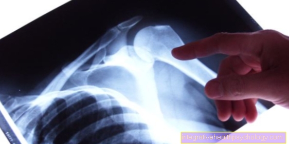
Which therapy achieves the best results in the long term can only be determined after looking at all of the information (Examination, X-ray, ultrasound, MRI, etc.) be assessed.
You can find me in:
- - your orthopedic surgeon
14
Directly to the online appointment arrangement
Unfortunately, it is currently only possible to make an appointment with private health insurers. I hope for your understanding!
Further information about myself can be found at
Symptoms
Indicative of the presence of a Meniscus lesion are some typical symptoms. In the foreground are of course those Pain. On the one hand, these can be Rotary motion and also in general under pressure occur or even get worse. Long-term damage often manifests itself in severe pain under stress.
At the same time as the pain, a Risk of entrapment and a Effusion from irritation exist. Occurs in the knee joint pain on the along the joint space runs, this is an indication of a lesion of the menisci. Other classic symptoms are Swelling, crepitus and the Restriction of movement. More precisely, they are Extension and flexion movement, so extension and flexion are hardly or not at all possible. Furthermore, it can become a Blockage in the knee joint come as a result of a meniscus tear. If the torn or torn part of the meniscus becomes trapped in the joint, this event triggers severe pain and a joint blockage. This is also considered a symptom Presence of a positive function test for the menisci.
Clinical tests

With the help of special function tests Is it possible, relatively safe to diagnose a meniscus lesion. Due to the Variety of tests is a clear differentiation regarding the inner or outer meniscus, localization and Type of lesion possible. A positive test is indeed indicative on a meniscus lesion, however accurate diagnosis only by combination the various function tests. It is the basis To put menisci under stress through pressure, shear or tension. If that leads to a Release of pain comes one speaks of one positive test; otherwise a test will be negative. As mentioned, there are countless functional tests available for assessing a meniscus lesion, which are explained below.
Apley compression test (grinding test)
The patient lies on his stomach and has a Knees bent 90 degrees. The examiner fixed now with one hand or the leg Thigh of the patient. Simultaneously rotates he now does that with the other hand leg of the patient once negative pressure and once under train. It comes with the Internal rotation to pain is the External meniscus damaged, however, hurts the External rotation lies a Medial meniscus damage in front.
Böhler-Krömer test
This test is also called a Abduction and adduction stress test called. The examiner stabilized with one hand Thigh of patient lying on their back and with the other hand it includes the ankle region. To the Medial meniscus to test engages the upper hand to the Inside of the thigh or the knee and the lower hand to the Outer ankle. Now bends and stretches the examiner holds the leg while he is there simultaneously adducted, so under Varus stress puts. To the External meniscus need to test the Grasp each handon the outside of the thigh and on the inside of the ankle. Then one can Valgus stress be exercised with simultaneous flexion and extension, i.e. an adduction movement. Of the Compression pressure can be used in both investigation steps Pain trigger in the appropriate meniscus and indicate its lesion.
Bragard test
From the Supine position should Knee joint in a 90 ° position be. The examiner now includes the foot with one hand and felt with the other hand medial (to the middle of the body) and the lateral (to the side of the body) Joint space. Now he rotates the leg once outwards and inwards and then changes from the flexion to the extension position. If the Inner meniscus damaged is, occur in the Bragard test on the one hand Pain from palpation of the medial joint space on and on the other at the External rotation. The transition to the extended position with further external rotation usually makes the pain worse. In the External meniscus lesion it behaves exactly vice versa: Pain to the lateral joint space and at Internal rotation.
Cabot test
This test checks whether the patient will be Extend the knee against the resistance of the examiner can without causing pain. The starting position provides that the Patient lies on his back and the affected Leg rolls over slightlyby placing the foot on the lower leg of the healthy side. The examiner includes the lower leg and palpated at the same time lateral joint space. Now he is asked that the patient Straighten leg while the examiner holds back slightly. Depending on how severe the pain is, the patient may not be able to fully extend the leg. Of the pain occurs at a External meniscus lesion at the external joint space on and can partly pull into the posterior joint space area.
Childress sign
The patient takes one deep squatting position a so that the Heels already that Touch buttocks and should then move in the duck path. Is there a Meniscus lesion, more precisely a lesion of the posterior horn of the menisci, in front of the patient unable to duck, because he strong pain felt at maximum flexion and at the time of extension. The pain can be from one "Crackling - or snap-like" sound be accompanied, since at this moment there is an entrapment of the menisci.
McMurray test (Fouche sign)
Here lies the Patient on his back and bends his leg both in the knee and in the hip joint maximum. Of the Examiner now covers the leg of the patient and leads a Internal and external rotation by. The examiner continues the two rotations while doing this Extends the patient's leg to a right-angled knee joint position. Pain in the External rotation have a Medial meniscus lesion as a cause, pain Internal rotation are through one External meniscus damage justified. It comes to one during the stretching "Snap-like" sound the lesion is more in the middle area of the meniscus. However, if the snapping sound occurs in the maximum flexion position, the posterior part of the meniscus is more likely to be damaged.
Anderson compression test
The examiner clamps the leg of the patient lying on his back under his arm and palpated with your free hand Joint space. After that bends and stretches he that knee of the patient. During the diffraction he also practices one Valgus stress, in the Stretching a varus stress out. Pain can be used with this function test on a Longitudinal or flap tear provoke the meniscus.
Remember test
The memory test checks them Rotational ability. The patient is prompted to stand on the leg of the sick side and this easy to bend. Then he should Lift the other leg slightly and the Thigh once turn outwards and inwards once. This limits internal and external rotation of the lower leg on the diseased standing leg, which the examiner can fix on the floor for assistance. Solves the External rotation of the thigh Pain off, this points to one External meniscus damage because the movement of the external rotation on the healthy thigh corresponds to an internal rotation on the lower leg of the sick leg.The opposite applies: Internal rotation pain of the thigh are indicative of one Medial meniscus damage.
The pain symptoms are particularly pronounced, since in this test the entire body weight rests on the knee and thus the menisci, so that the axial compression or the compression pressure is very high.
Payr sign
For this test, the patient must be on the examination table Cross-legged position take in. Of the Examiner presses his knees, which are currently in an externally rotated and bent position, now moderately strong towards the substrate. Will the Applying pressure in the medial joint space as painful felt, the patient probably has one Meniscal lesion in the posterior area.
Steinmann I.
The Steinmann I test aims at one Rotational pain trigger. The patient lies on his back and bend the knee about 30 °. The examiner grips the lower leg with one hand and the heel with the other, from where he takes one Internal rotation and then a External rotation performs. Typically speaking External rotation pain for one Damage to the medial meniscus and at the Internal rotation for one Damage to the external meniscus.
Steinmann II
In the second Steinmann test, the patient is also lying down on the back. The examiner tries through that Palpation of the medial and lateral joint space one pain of the respective meniscus trigger A medial pain speaks for one Medial meniscus lesion. It is important that the pain point can migrate: When flexing, the pain moves backwards in the affected joint space and forwards when stretching. also rotates the examiner that Patient's leg inwards and outwards while at the same time one axial compression (i.e. pressure from below against the knee joint). External rotation pain speaking for Medial meniscus lesions and Internal rotation pain For External meniscus lesions.
Thessaly test
In this test the patient stands barefoot on one leg. In order to be able to keep the balance, the examiner holds the patient's outstretched arms. Now that's supposed to Support leg bent by 5 ° and then the Upper body rotated 3 times outwards and inwards become. First the Performed on the healthy leg, then only on the sick leg. Then the patient should do it again, albeit with 20 ° flexion (Flexion) in the knee. Pain that appear in the joint space in this test speak for one Meniscus lesion on the corresponding aching leg. In addition, the Thessaly test can Joint blockage provoked become.
Turner sign
At the Inside of the knee joint is in the presence of a Medial meniscus lesion a hypersensitive (hyperesthetic) Skin area. Mechanical and thermal irritation can test Turner's sign as positive and indicate a meniscus lesion. However, the reason for the hypersensitivity can also be the chronic irritation of the supplying Nerve, a branch of the saphenous nerve (R. infrapatellaris nervi sapheni).
Rotational compression test according to Pässler
The patient takes on a chair Space. The examiner stuck yourself that affected leg between his own legs approximately at the level of the knee joints and felt then with both thumbs medial joint space. To now one Internal and external rotation In order to be able to perform, the examiner has to make a kind of circular motion; the leg continues to remain wedged between its own. The rotation test solves a Medial meniscus lesion Pain at the medial joint space off, at one External meniscus lesion creates pain on the outside of the knee joint, i.e. on the lateral joint space.
Tschaklin sign
The Tschaklin sign is not really a function test. The Tschaklin sign is considered positiveif on the one hand by a older meniscus lesion to tissue shrinkage (atrophy) of large thigh muscle (Quadriceps muscle) or if, on the other hand, the vastus medialis muscle is atrophied due to a medial meniscus lesion and another muscle, the sartorius muscle, increases its tone for compensatory reasons.