MRI for a meniscus tear
introduction
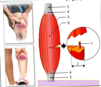
The Menisci are cartilaginous structures of the Knee joint. They are between the articulating bones, so between the Thigh bone (lat. femur) and the lower leg bone (Latin tibia).
The menisci serve to create a better contact between the two bones and compensate for the incongruence due to their different shape and curvature. In addition enlarge she the force-absorbing surface in the knee joint and thus ensure a better distribution of joint pressure.
There is an inner and an outer meniscus. Both are on the tibial plateau in a certain region, the so-called "Area intercondylaris anteriores and posteriores“Anchored on the bone.
Of the Medial meniscus is also connected to the outer ligament (collateral ligament) of the knee joint so that it extends far less agile is than the outer meniscus. Both have one crescent shape; the outer meniscus even represents an almost closed ring.
Meniscal tear
Like any structure, the menisci as an important part of the knee joint can be damaged.
The Meniscal tear as the most common injury. This is understood to be an interruption in the continuity of the cartilaginous structure. The causes are usually traumatic events or degenerative processes.
Strong shear forces, twisting or dislocating the knee, falls, and an abrupt stop to movement can all lead to a Meniscal tear to lead. Especially in old age, but possibly also at a young age, signs of wear and tear and overuse make a meniscus tear more likely.
Of the Medial meniscus is due to its reduced mobility due to the additional attachment to the Inner band from the knee joint more often affected.
The posterior third (posterior horn of the medial meniscus) usually ruptures. In addition to the localization of a meniscus tear, one can different crack shapes classify (Transverse, Along- and Meniscus tear like a handle basket).

I would be happy to advise you!
Who am I?
My name is I am a specialist in orthopedics and the founder of .
Various television programs and print media report regularly about my work. On HR television you can see me every 6 weeks live on "Hallo Hessen".
But now enough is indicated ;-)
The knee joint is one of the joints with the greatest stress.
Therefore, the treatment of the knee joint (e.g. meniscus tear, cartilage damage, cruciate ligament damage, runner's knee, etc.) requires a lot of experience.
I treat a wide variety of knee diseases in a conservative way.
The aim of any treatment is treatment without surgery.
Which therapy achieves the best results in the long term can only be determined after looking at all of the information (Examination, X-ray, ultrasound, MRI, etc.) be assessed.
You can find me in:
- - your orthopedic surgeon
14
Directly to the online appointment arrangement
Unfortunately, it is currently only possible to make an appointment with private health insurers. I hope for your understanding!
Further information about myself can be found at
Diagnosis
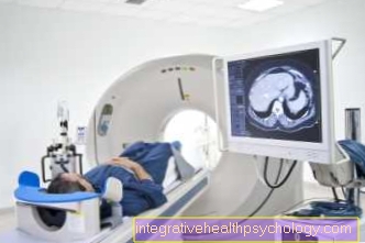
If a meniscus tear is suspected, a clinical examination with special tests comes first. So-called Provocation test enable the detection and differentiation of inner and / or outer meniscus tears through compression and rotational loading. Other methods would be Computed Tomography (CT; layered x-ray), ultrasound and, as an invasive method, knee arthroscopy. With chronic meniscus lesions, a Arthroscopy be indexed. The acute / fresh meniscus tear is far more common. That is true MRI (Magnetic resonance imaging) as the means of choice for imaging procedures.
Diagnostics of choice - MRI
With the help of Magnetic resonance imaging (or Magnetic resonance imaging) a meniscus tear can be assessed more precisely in terms of its shape and extent.
The principle of MRI is based on the magnetic property of individual atomic nuclei in our body, which each have a certain characteristic intrinsic angular momentum. The exact function is very complex - simply put, a computer can record and evaluate the impulses, so that in the end a three-dimensional image arises.
The MRT is a 3D recording process that can show meniscus tears in every spatial plane. Through the use of magnetic fields and radio waves, the MRI image offers the possibility of different things Image contrasts. The MRI image is weighted depending on which tissue can be assessed. The basis for this are the Hydrogen atomsthat are everywhere in our body occur, however different density and often in different organs.
For example, in some weights the muscles or menisci appear darker and fluids appear lighter (T2-weighted image), in other images, structures rich in fat are lighter (T1-weighted image).
Using this principle of different contrasting different structures can be clearly delimited from one another on an MRT image.
In general, the MRI has the advantage that it can be compared to x-rays no radiation exposure (by ionizing rays) there.
It is also very well suited for displaying soft tissue, including the menisci and other structures (ligaments, cartilage and synovial membrane) of the knee joint, as the smallest lesions and irritation can be recognized.
The MRI is therefore now considered the gold standard in the diagnosis of meniscus tears.
MRI procedure for a meniscus tear
Nowadays, the MRI is performed by radiologists and medical-technical radiology assistants (MTRA) either in the clinic itself or, in the case of less acute concerns, in radiological studies. It is very important to inform the patient about the subsequent examination in advance, as the doctor may ask about possible contraindications in such a conversation.
As already mentioned, an MRI works among other things with magnetic fields so that metallic objects attracted to the device and can be heated. This represents a potential risk of injury to the patient, but it can also severely damage the MRI machine itself. Wear Patients, for example a pacemaker, are you allowed to no MRI scan do.
Jewelry, glasses, watches and other metallic objects must be removed beforehand. During the examination, the radiologist and the technologist leave the room in which the MRI machine is located.
However, you can monitor the patient from a separate room through a glass window. In addition, there is an intercom system for mutual communication and, in the event of an emergency, a safety bell that can be operated by the patient.
Often times, patients have Claustrophobiabecause the device is very narrow and tight.
Please also read our topic: MRI for claustrophobia
To clarify a meniscus tear, however, the head is usually in the open. It is important to know, however, that the MRI examination is completely painless and you usually do not notice anything. The only thing that you can hear are the loud noises emanating from the MRT machine (“knocking noises” / clacking / “rattling”). Earmuffs or headphones with music can be worn here for cushioning and distraction.
A meniscus tear does not take quite as long to perform as more complicated or extensive analyzes.
Duration
The examination takes a maximum of 20 minutes. There is also the time to clarify and prepare and possibly waiting time on site.
The use of contrast media
The MRI examination is done nothing inevitably using contrast media. The use of certain contrast media (contrast media) depends entirely on the question.
The idea behind this is that some structures are displayed in similar gray levels without contrast agent, which can be a hindrance to the diagnosis. The administration of a contrast agent can a better color gradation can be achieved, since different tissue structures absorb and enrich the contrast medium to different degrees, and this leads to increased contrast. Areas of tissue that absorb a KM particularly well are brighter. There is also another difference between “whitening / lightening” and “darker / blackening” contrast media. However, it becomes an area despite KM gift contrary to expectations not brighter, can this an indication of it be that the appropriate structure not well supplied with blood is.
The KM will namely via an arm vein injected and should be distributed throughout the body via the bloodstream. The menisci are subdivided into 3 zones depending on the blood flow so that a changed blood flow can be recognized in the MRI.
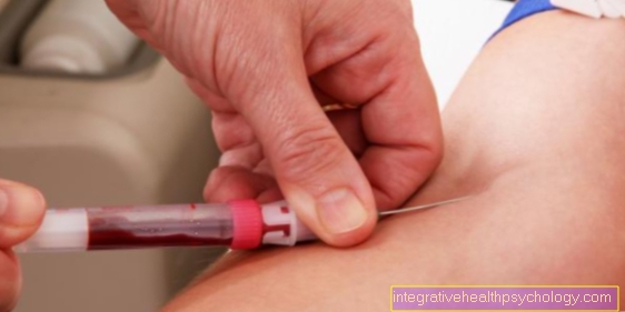
A very commonly used one Contrast media is this Gadolinium. There is no addition of gadolinium toxic and can accumulate in organs such as the liver, bones or spleen. Hence it is called an essential acid DTPA to add. The elimination of the contrast medium then takes place via the kidney. Occasionally, there may be intolerance to the contrast agent. Patients who have a tendency to allergies should therefore inform their attending physician in any case.
Contraindications
The general ones apply Contraindications as with any MRI. Implants like Pacemaker or Cochlear implants are considered a contraindication. Endoprostheses like Hip TEPs (Total replacement of the hip joint) and screw or plate material are usually made of titanium, which is compatible with MRI, but can heat up during a long examination and can lead to artifacts, i.e. disturbances in the image.
Pregnant women should be in early pregnancy not getting an MRIas it is not known for sure whether this can cause harm to the fetus.
Other metal parts, for example Metal splinters after accidents, are also considered a contraindication to the examination.
For patients with fear of tight spaces (claustrophobia), an MRI scan of the knee should be tolerable, as the head and most of the body are not in the MRI tube.
The administration of contrast media can very rarely lead to allergic reactions and contrast media should not be given if kidney and liver values are poor.
Alternatives to MRI for meniscus tears
Even if the MRI is the gold standard in diagnosing a meniscus tear, this is true CT (Computed Tomography) as a possible alternative. This is an X-ray process (ionizing radiation), which Compared to pure X-ray, also show soft tissue can
The difference between MRI and CT is the radiation exposure, which is often interpreted as a disadvantage of CT. In addition, the CT is inferior to the MRI in the accuracy of the soft tissue representation, so that it is used less often than the MRI to assess a meniscus tear. However, computer tomography is very suitable for detecting broken bones or calcifications. It is also often used to plan prosthetic treatments.
costs
The cost of an MRI scan for a meniscus tear is the same as that of the knee joint. Private patients and self-payers at least € 139.89. A maximum of € 349.72 can be billed.
In addition to the costs for the MRI examination, there may be costs for advice, contrast media and further exposures in a different joint position, so that costs of more than € 600 can arise in the end.
As a privately insured person, always find out exactly what the costs will be in advance.
In the case of statutory health insurance patients, the radiologist may charge the Association of Statutory Health Insurance Physicians € 124.60 for the pure MRI examination of the knee joint. Billing takes place directly between the radiologist and the Association of Statutory Health Insurance Physicians.
Read more about the implementation and indication of a knee MRI under: MRI of the knee


.jpg)