CT of the lungs
definition
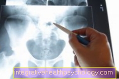
A commonly used imaging technique to visualize the lungs is computed tomography (CT). This is a special X-ray examination in which several cross-sections of the body are recorded and combined to form a three-dimensional image with very high resolution. The imaging is done with the help of X-rays, which are absorbed to different degrees by the various tissues of the body.
A so-called CT chest is made to visualize the lungs. This is an image of the chest (lungs and heart). It is often used as a supplement to conventional X-ray images. A particularly high-resolution computer tomography (HRCT) method is used for individual lung diseases.

Indications for a CT scan of the lungs
Computed tomography of the lungs is used in the diagnosis of many diseases. Compared to conventional X-ray images, it is characterized by a significantly higher resolution and a three-dimensional display, which means that very fine structures in the lung tissue can also be displayed.
The chest CT is performed particularly often to identify, monitor the progress and follow-up of tumors and metastases in the lung area. In addition, numerous inflammatory diseases of the lungs can be shown well in the CT chest.
In addition to classic pneumonia, chronic obstructive pulmonary diseases (COPD) can also be diagnosed. The CT chest is also suitable for displaying vascular changes in the area of the large pulmonary arteries. Vascular occlusions (e.g. in the case of a pulmonary embolism) and vascular changes (e.g. due to atherosclerosis or aneurysms) can be clearly identified with the aid of computed tomography.
Last but not least, the CT thorax is also used to plan major operations in the thoracic area, as structures relevant to the operation are displayed very precisely.
Depending on the question, the administration of an iodine-containing contrast agent may be necessary in order to be able to better differentiate the individual tissue structures from one another in terms of color.
Also read: CT of the chest / upper body
Pulmonary embolism
Pulmonary embolism is a blockage of one or more pulmonary arteries. This often happens through a flooded thrombus, which mainly comes from the area of the deep leg veins. As a result, the lung tissue is less supplied with blood and the right heart is more stressed.
A CT-controlled display of the vessels (CT angiography) is suitable for visualizing a pulmonary embolism. For this purpose, the patient is administered an iodine-containing contrast medium intravenously. This allows the thrombus to be clearly differentiated from the otherwise well-perfused vessel in the CT images. In addition to confirming the diagnosis, blood is often taken and the D-dimer concentration in the blood determined.
You might also be interested in: This is how you can recognize a pulmonary embolism
lung infection
Inflammation of the lungs (pneumonia) can manifest itself in different ways in the area of the lungs. Both the air-filled alveolar space (alveolar pneumonia) and the connective tissue of the lungs located in between (interstitial pneumonia) can be affected. Depending on the age and the cause of the infection, a wide range of pathogens (bacteria and viruses) can be responsible for the development of pneumonia.
The main criterion in the diagnosis of pneumonia is a newly occurring infiltrate that has been detected in the X-ray. This appears as a white shadow in the area of the air-filled (black) alveolar space. If the X-ray findings are unclear, a CT chest can also be made to confirm the diagnosis.
Read more on the topic: How do I recognize pneumonia?
Lung cancer
Computed tomography is also used regularly in the identification, progress and follow-up control of lung tumors and metastases. Fuzzy nodules of the lungs appear suspiciously, as white shadows.
Depending on the type of tumor, these can be located in different parts of the lungs. Basically, every unclear nodule in patients over 40 years of age is assessed as lung cancer until proven otherwise.
A contrast agent is often administered during the course of the CT diagnosis to better delimit and assess the blood flow to the tumor. In order to confirm the diagnosis, a puncture of the tumor (lung biopsy) is also carried out with the help of a CT in order to be able to analyze the tumor tissue.
You might also be interested in: Diagnosis of lung cancer
COPD
Chronic obstructive pulmonary disease (COPD) causes inflammation of the small airways (chronic obstructive bronchitis), causing them to become increasingly blocked and the lungs to overinflate (emphysema). As a result, breathing is significantly restricted.
There are several ways to diagnose COPD. In addition to a routine test of lung function, a conventional chest X-ray is used to show the overinflation of the lungs.
To better assess the localization and distribution of the emphysema, a CT chest be performed.
Read more on the topic: Diagnosing COPD
Pulmonary fibrosis
Lung fibrosis is the final stretch of many different lung diseases. This leads to a significant increase in the connective tissue within the lungs, which makes breathing difficult for the patient. This is often caused by chronic inflammation in the lung area.
Lung fibrosis is also diagnosed by testing lung function and imaging using X-rays. A conventional chest X-ray is often supplemented by a CT chest.
More information is available here: The pulmonary fibrosis
Preparation of the CT thorax
Before performing a chest CT scan to visualize the lungs, there is always a consultation with a doctor.
This preliminary discussion serves to explain the benefits and risks of the investigation. The patient should be informed of the radiation exposure during imaging.
If a contrast agent is planned to be administered, the doctor must be informed about the intake of medication, known intolerances and allergies as well as existing pre-existing diseases (e.g. liver and kidney diseases).
In most practices and clinics, the patient should be asked for Be sober for 6 hours to ensure better quality images.
Procedure of the CT thorax
During the imaging, the patient is on a kind of table that is increasingly driven into the CT device in the course of the examination. The X-ray tube and the opposite detector system rotate around the patient during the course of the examination, so that the individual layers of the body can be recorded. Compared to an MRI machine, the CT tube is so short that the patient can look out of the tube during the entire examination and there is usually no fear of claustrophobia. Nevertheless, a sedative can be administered before the examination.
No other person is in the room during imaging in the CT device. The patient can call the staff at any time via an intercom. To examine the lungs, the patient must hold their breath for about 10 to 20 seconds at regular intervals so that the images recorded are of good quality and the lungs are displayed in their expanded state.
Depending on the question, the patient is given a contrast medium approximately 1 hour before the examination.
Do you always need contrast media?
The administration of an iodine-containing contrast medium depends on the question to be examined. The contrast agent is used to better represent structures with blood flow. In particular, inflammations and tumors turn very white after administration of the contrast agent and can be distinguished from their surroundings. In addition, the contrast agent is also suitable for depicting a pulmonary embolism, since it clearly distinguishes the color of the blood from that of the thrombus.
Various side effects can occur when a contrast agent is administered. In a preliminary talk, the doctor should be informed about known intolerances, allergies and previous illnesses.
Read more on the topic: Contrast media
Diagnosis of a CT chest
The images taken during the examination are usually viewed by a doctor immediately afterwards. As a rule, they can communicate the first results immediately. However, since a total of up to 100 images can be recorded with CT imaging, the doctor only creates a written report after a detailed evaluation. These findings are usually passed on to the treating doctor within a few days.
Duration of the CT examination
The CT examination is an imaging diagnosis that can be carried out quickly and easily. For this reason, it is often preferred to an MRI scan. Computed tomography should be performed much faster, especially in emergency situations.
Depending on the question, a CT chest to examine the lungs takes between 5 and 30 minutes. Usually the duration is about 10 to 20 minutes. However, the administration of a contrast agent can add a few minutes to the imaging time.
Radiation exposure for a CT chest
In computed tomography imaging is done with the help of X-rays that are harmful to humans. Compared to conventional X-ray diagnostics, the patient is exposed to significantly higher radiation doses during the CT examination. However, the risk of damage to health from this radiation exposure is low. The further development of CT devices has made it possible to reduce the radiation dose and the duration of the examination to be shortened.
The exact radiation exposure of an examination depends on the number and thickness of the slices and the structure of the tissue to be examined. The average radiation exposure for a CT chest is approximately 6 to 10 milli-Sieverts (mSv). In comparison, the annual average radiation exposure for people living in Germany is 2.1 mSv.
Read more on the topic: Radiation exposure in computed tomography
Cost of a CT scan of the lungs
The costs of a CT examination of the lungs are covered by the statutory health insurances for clinically indicated questions for patients with statutory health insurance.
The price for CT imaging in the neck and / or thorax area for self-payers and private patients is € 241.31 according to the fee schedule for doctors (GOÄ).
A discussion in advance and a possible debriefing can make the total costs a little higher.













.jpg)

