MRI of the sacroiliac joint

introduction
An MRI, i.e. magnetic resonance tomography, is an imaging process in which the patient is driven in an elongated tube and sectional images of the body are created. In contrast to a CT or X-ray, MRI does not work with X-rays, but with a magnetic field that stimulates hydrogen nuclei in the body's cells.
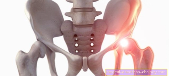
The joint between the pelvis and the spine is called the SI joint (sacroiliac joint). There is often pain there. The anatomy and disease-related changes can be determined through the MRI cross-sectional images.
Indications for an MRI of the sacroiliac joint
An MRI is often used to diagnose pain in the lower back or pelvis, as it provides reliable images of the soft tissues, i.e. muscles, tendons, adipose tissue, organs and bones.
Do you have lower back or pelvic pain? - then read our articles on this Lower back pain or Pelvic pain
The MRI can diagnose diagnoses such as sacroiliitis or SI joint arthrosis.
MRI is not always the first choice; CT (computed tomography) is more often performed.
However, since an MRI works without harmful X-rays, it is often used on children or pregnant women.
Also, because an MRI has a better representation of the muscles and soft tissue than a CT, the intervertebral discs, muscles, and spinal cord can be better visualized.
Sacroiliitis
Sacroiliitis is an inflammation of the sacroiliac joint. It often occurs in the context of other, especially rheumatic, diseases, such as M. Bechterew, M. Behcet, M. Reiter (so-called Reiter syndrome).
The SI joint connects the pelvic vane (os ilium) with the lower part of the spine, the sacrum (os sacrum).
In the case of sacrolitis, among other things, pain in the lower back, which occurs mainly at night and in the morning after getting up.
As a rule, there is an improvement in the course of the morning (starting pain). Often both sides are affected.
The diagnosis is made with the help of an MRI of the ISG. Treatment is with anti-inflammatory and pain reliever drugs, as well as physiotherapy.
Do you want to do something about your back pain yourself? - Then read our article Exercises for back pain
SI joint osteoarthritis
Osteoarthritis describes a degenerative change in the articular cartilage that has occurred due to wear and tear.
This usually occurs in older people and can have different causes, in most cases it arises from long-term excessive or improper stress.
A pelvic inclination can also be the cause. The consequences are wear and tear on the joint surface and the resulting pain in the lower back, as well as restricted mobility.
These increase over time and often worsen with exertion. The joint surface and the articular cartilage can be assessed well in the MRI. A reduced joint space and protruding bones are signs of advanced osteoarthritis.
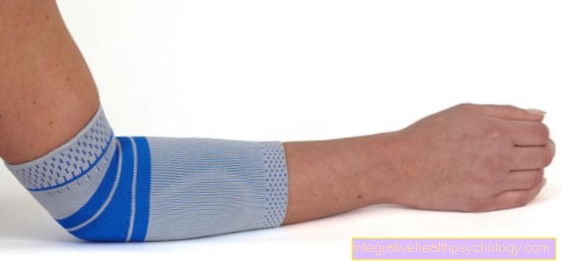
SI joint arthrosis can be treated conservatively with medication or bandages, or surgically.
Would you like more detailed information about what can be done against osteoarthritis? - Then read our article Osteoarthritis therapy
Procedure for the MRI examination of the sacroiliac joint
The process of an MRI examination begins with its preparation.
In preparation, the doctor first informs you about the upcoming examination. There you will also find information about possible risks of an MRI examination.
It is not necessary to be sober before the examination.
In some cases, contrast agent is given beforehand through the vein. Since the MRI works with the help of a strong magnetic field, it is very important to remove all metal-containing parts on the body before entering the room.
This includes piercings, jewelry, cell phones, credit cards, etc. Otherwise, this can have dire consequences.
The MRI is an elongated tube with a hole in the middle through which a couch goes. The patient is completely or only partially driven into the tube on this couch.
When the MRI is on, it is usually very noisy, so patients always wear hearing protection and headphones.
The examiner who is outside the room can also communicate with the patient via these headphones.
In most cases, the tube is very narrow and you should move as little as possible. This is a big problem, especially for patients with claustrophobia. It is possible to give sedatives in advance.
Do you suffer from claustrophobia and still need to do an MRI? - Then read our articlel MRI for claustrophobia - What are the options?
Is contrast agent necessary for a sacroiliac joint MRI?
With an MRI of the ISG, the administration of contrast media is usually not necessary.
Contrast media is used in particular when the examination is indicated to visualize the soft tissue.
The contrast agent collects in organs and muscles and can therefore help with the diagnosis.
However, the administration of contrast media also involves risks, as allergies and kidney damage can occur.
If the ISG is displayed alone, contrast agent is not required, since here mainly the bones and joint surfaces are examined.
Duration of the MRI scan of the sacroiliac joint
The duration of an MRI examination of the ISG is usually around 15-25 minutes. It depends on what is to be examined.
In addition, there is the preparation time, i.e. the undressing of the patient, the positioning and the evaluation of the images. A total of at least one hour should be planned for an MRI appointment.
Do I have to put my head in the tube?
During an MRI examination of the ISG, you usually do not have to put your head into the tube.
However, this depends on the size of the patient. The patient is driven into the MRI tube with his feet first on the couch until his entire pelvis is in the tube.
Evaluation of an MRI examination of the sacroiliac joint
In most cases, the evaluation is carried out by an experienced radiologist.
During the evaluation, all sectional images are viewed in 3 levels on the computer. A possible change in the ISG can be precisely localized through the various levels.
With the ISG, the doctor assesses the joint surface and the bone substance. For example, is there a narrowing of the joint space? Are there any cysts visible?
In addition, the whole picture is always considered, i.e. adjacent structures that have nothing to do with the diagnosis are also assessed.
If the patient has already had an MRI in the past, the current one is compared with this and possible changes are noted.
Cost of an MRI scan of the sacroiliac joint
The exact costs of an MRI of the ISG cannot be precisely determined, as they depend on various factors.
On the one hand, the patient's health insurance is decisive, i.e. whether they are privately or legally insured.
In addition, other factors can be factored into the costs: How many markers were made? Was contrast media used? How long did the investigation take?
As a rule, an MRI examination of the ISG costs between € 400 and € 800. In most cases, however, these costs are covered by the health insurance company if there is a medical indication.
What are the alternatives to the sacroiliac joint MRI?
If there are contraindications for performing an MRI, for example claustrophobia or a defibrillator, the question of an alternative arises.
Do you suffer from claustrophobia and still need to do an MRI? - Then read our articlel MRI for claustrophobia - What are the options?
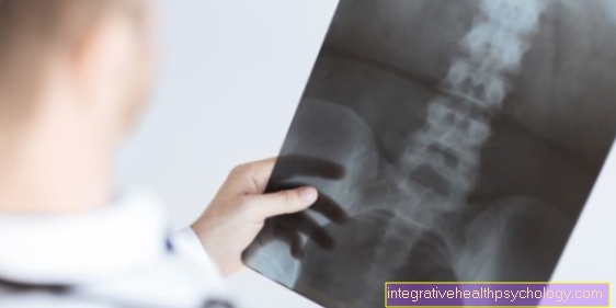
This depends on the question and the indication. If there is pain in the back or pelvis, an X-ray can also be taken.
This allows the bone structures to be examined and osteoarthritis diagnosed. However, this means that the soft tissues cannot be assessed.
Another alternative is CT (computed tomography). This also works with ionizing radiation, but this allows the bone structures to be better represented. It also provides sectional images and can help in finding a diagnosis.
How well can you see inflammation on an MRI scan of the sacroiliac joint?
Inflammation can be seen very well in the MRI.
Inflammation usually appears as a lightening in the soft tissues or in the joint space. This lightening can be compared with the opposite side.
In addition, the administration of contrast media can be helpful, since inflammatory changes lead to increased blood flow and thus more contrast media accumulates.
Can you do an MRI examination of the lumbar spine and the SI joint?
An MRI of the lumbar spine (lumbar spine) and sacroiliac joint (SIJ) can be done together.
To do this, the patient is simply moved a little further into the tube on the couch. In the case of a sole ISG exposure, the lumbar spine is usually partially shown.
If the lumbar spine is also to be examined, sectional images are made up to the lower thoracic spine.
This is how you can assess both the lumbar spine and the SIJ. This is particularly useful If you want to rule out a herniated disc of the lumbar spine as the cause of the pain.
Is an ISG blockage visible on the MRI?
An ISG blockage is a blockage in the area of the joint surfaces. The various joint parts can no longer move without problems.
This leads to pain and restricted mobility. In addition, there can be discomfort and tingling on the feet.
As a rule, the blockage can be quickly cleared with targeted movements and no MRI examination is required.
In any case, it can only be diagnosed to a limited extent in the MRI. Only if the blockage is very severe is it visible on the MRI. Possibly. A build-up of fluid may be visible around the obstruction.