Tibialis posterior muscle
definition
The tibialis posterior muscle is a skeletal muscle that lies in the area of the calf and extends with its tendon around the inner ankle to the sole of the foot. It is officially part of the lower leg muscles, which can still be divided into deep and superficial muscles. The tibialis posterior muscle belongs to the deep muscle group.
It arises on the two lower leg bones and lies wedged between the two other muscles of the deep lower leg group. Due to its position and course, contraction results in an extension of the upper ankle joint, a supination of the lower ankle joint and a tension in the longitudinal and transverse arches of the foot. The nerve supply is provided by the tibial nerve.

Anatomy of the tibialis posterior muscle
Anatomically correct, the muscle is counted as part of the deep flexor group. These three muscles all originate in one or both bones of the lower leg. The tibialis posterior muscle originates from the back of the tibia and fibula. It also arises from a very tight membrane (Membrana interossea cruris) that lies between these two bones.
On the lower leg, it lies between the flexor digitorum longus and the flexor hallucis longus muscles. From here, the insertion tendon passes under the insertion tendon of the flexor digitorum longus muscle below the inner ankle over the upper and lower ankle joint to the sole of the foot. Here the tendon attaches to several bones of the tarsus and metatarsus.
In addition to the superficial flexor group, the tibial nerve also supplies the entire muscles of the deep flexor group with signals. Due to its anatomically deep position in relation to the body surface, injuries are rare.
Posterior tibial tendon
The tendon of the tibialis posterior muscle, like all tendons in the body that are connected to the muscles, consists of very tight collagen fibers that have a high tensile strength. The tendon begins at the lower end of the tibialis posterior muscle. However, the transition cannot be precisely determined.
In the distal part of the lower leg there is a junction with the insertion tendon of the flexor digitorum longus muscle. The insertion tendon of this muscle crosses the insertion tendon of the M. tibialis posterior (,, dig over tib ''). However, this is not a hindrance to everyday life.
From there, the insertion tendons of the tibialis posterior muscle pass through the malleolar canal. This is located below the middle ankle. Here, all the tendons are encased in a tendon sheath to minimize friction between the individual tendons. The tendon is attached to the navicular bone, the sphenoid bones and the metatarsal bones.
Further information on the Tibialis Posterior Tendon and its diseases can be found at: Tibialis posterior tendon
Function of the tibialis posterior muscle
The functions of the muscle result mainly from the position and course of the muscle and its attachment tendon. As already described, the attachment tendon runs towards the back of the upper ankle joint towards the foot and attaches to the underside of the bones there. This results in a contraction with the accompanying shortening of the muscle belly, a movement of the tip of the foot away from the body, as when walking on tiptoe. This is also known as plantar flexion.
Since the insertion tendon also passes the lower ankle joint in the middle of the ankle, when the muscle contracts, the sole of the foot is raised towards the middle. This process is also known as supination or inversion.
An isolated movement of the upper or lower ankle joint when using the M. tibialis posterior is usually not possible, as the muscle always performs both functions when it contracts. Only the interaction of several muscles allows an isolated plantar flexion or an isolated supination.
Since a person's weight always creates tension on the attachment tendon, it also tensions the two arches of the foot. The tendon runs on the sole of the foot both slightly transversely and slightly obliquely in relation to the body axes and can thus stabilize both arches.
Diseases of the tibialis posterior muscle
Tendonitis of the tibialis posterior muscle
Inflammation of the tendon can manifest itself in several ways and have a variety of causes. In the case of inflammation, the tendon sheath in the malleolar canal is usually also affected.
The most common cause of inflammation of the tibialis posterior tendon is overexertion. Doing this too often can be the only decisive factor. However, even a minimal incorrect posture during repetitive movements can cause minor injuries to the tendon and tendon sheath. Typically, such an overload occurs in athletes. However, prolonged walking uphill can also be a cause.
Other possible causes are infection or an immunological reaction in the body. Such an inflammation becomes noticeable through permanent pain in the muscle area. There may also be swelling and persistent pulling. Later, the muscle strength decreases, making it difficult to stand on tiptoe.
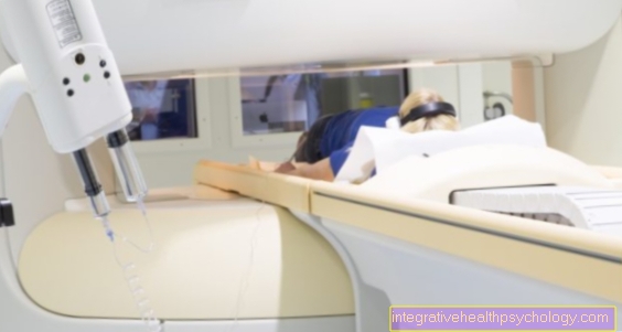
Furthermore, a misalignment of the foot or the tendon can tear if the inflammation persists, so that the clinical picture should always be treated. The diagnosis is usually based on a clinical examination. If necessary, imaging such as MRI can be used. An anti-inflammatory drug (NSAIDs) can be taken.
More detailed information on this topic can be found at: Tendinitis in the leg and tendinitis of the tibialis posterior tendon
Torn tibialis posterior tendon
Torn tendons are usually caused by chronic tissue degeneration. However, the rupture is perceived as an acute, very painful event. Furthermore, the tendon may tear as a result of inflammation of the tendon. A traumatic event such as a broken bone or a cut can also lead to a rupture.
Since degeneration is the most common cause, tendons tend not to tear in young athletes, but rather in older adults. Typical signs of a tear are sudden pain in the inner ankle. Often this is the result of an ankle twist. It can also make it difficult to keep your balance and walk on uneven ground. Furthermore, a misalignment of the foot can occur.
The diagnosis is made on the basis of a clinical examination and treated conservatively with insoles for the shoes over several months. In serious cases, surgery can also be carried out to reattach the tendon ends together.
Are you more interested in this topic? Then read more under: Tibialis posterior tendon
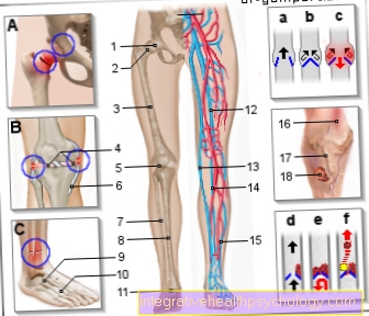
Figure torn hamstring
- Muscle fiber
of a skeletal muscle
Muscle fibra - Muscle fiber bundles -
Muscular Fasciculus - Tendon fibers -
Fibrae tendineae - Transition of muscle fibers
in tendon fibers -
Junctio myotendinea - Muscle fascia
(= Muscle skin) -
Fascia - Skeletal muscle -
Maecenas musculus osseus
You can find an overview of all Dr-Gumpert images at: medical illustrations
Recommendations from the editorial team
More information on other lower leg muscles:
- Clod muscle
- Anterior tibial muscle
- Twin calf muscle