Facial nerve
introduction
The facial nerve is one of the cranial nerves. There are a total of twelve nerves that originate in the brain and are responsible for various sensory perceptions, but also for movements. The facial nerve is the seventh of these cranial nerves. It is responsible for the movements of the facial muscles and, for the most part, for the sensation of taste. In addition, it also carries nerve fibers that supply glandular nerves.
If the nerve is damaged, what is known as facial paralysis occurs, in which the facial muscles (usually one-sided) can no longer be controlled arbitrarily.

Functions of the facial nerve
Since the facial nerve is composed of different parts, it also fulfills different tasks:
The motor fibers that are responsible for movements are mainly Facial muscles supplied, which are responsible for facial expressions. But muscles in the neck area are also innervated by the facial nerve, as is a small muscle in the ear (stapedius muscle) that is responsible for the Regulation of hearing in loud noises important is.
The sensitive parts that end in the skin and for that Sensation of touch and pain are responsible, take care of the Skin of the external ear canal and eardrum.
Other fibers (parasympathetic fibers) are responsible for the supply of various glands and end in the Oral salivary glands and the Lacrimal glands. The function of the glands is regulated via these fibers, and consequently their activation is preferred increased production of salivary secretions.
It also contains the facial nerve Flavor fibers (sensory fibers) that enable taste perception in the area of the front two thirds of the tongue.
Course of the nerve
The facial nerve has its origin in the lower, rear area of the brain, more precisely in the Brain stem. Here, its fibers arise from different nuclei located on both sides of the brain. There is therefore a facial nerve for both halves of the facewho takes care of one side.
The nerve runs both inside the skull (intracranial) and outside (extracranial) and leaves the brain in the area of the so-called cerebellar bridge angle. The nerve fibers then run inside the skull through the internal auditory canal and the temporal bone, whereby many branches for the supply of the muscles and the glands come off. Through the stylomastoid foramen, a hole in the skull behind the ear, the facial nerve emerges from the skull. He continues through the parotid gland and gives off smaller branches outside the skull to the muscles of the face and neck.
Irritation of the facial nerve
Permanent irritation of the facial nerve can trigger a facial spasm (so-called hemifacial spasm). A blood vessel often puts pressure on the nerves, causing damage to the insulating layer of the facial nerve. The excitability of the nerve is then increased and a state of permanent irritation occurs. This manifests itself in a one-sided cramping of the facial muscles, which usually only lasts for less than 1 second.
The causes of the irritation can be an aneurysm, i.e. the bulge on the side of a blood vessel, or, more rarely, brain tumors or multiple sclerosis.
Read more on this topic at: Facial palsy
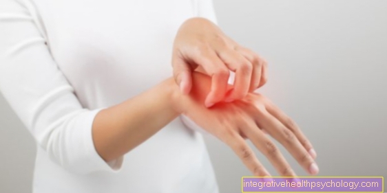
Pain
Pain caused by damage to the facial nerve is common a harbinger of facial palsy. Those affected usually complain of pain two to three days before the onset of hemiplegia in the area behind the ear. The lower jaw area can also be affected. If you experience very severe pain, you can use it, for example ASS (Aspirin®) are attempted to treat.
Facial paralysis (facial paralysis)

In the so-called facial paralysis or facial paralysis, one side of the facial muscles is paralyzed. Which side is affected depends on the cause of the paralysis and the location of the nerve damage.
A distinction is made between central and peripheral facial nerve palsy. In central paresis, the nerve is damaged in the brain and can be triggered by a stroke or a brain tumor. So the nerve itself is not damaged.
In peripheral facial paralysis, the damage affects the facial nerve itself. This can have various causes.
Facial paralysis manifests itself in incomplete eyelid closure on one side, drooping corner of the mouth, impaired taste sensation, hypersensitivity to loud noises and reduced tear and saliva production. Sensation is disturbed in a small area behind the ear. In the case of peripheral facial paralysis, in contrast to central paralysis, frowning is not possible. Due to the paralyzed facial muscles, there are often difficulties with word formation.
As a rule, one-sided facial paralysis disappears with the right treatment. Symptoms should no longer appear after six months at the latest. Permanent facial asymmetries can only be observed in a few cases, whereas in many people inconspicuous movements of the facial muscles remain when talking.
causes
Often are Circulatory disorders of the nerve responsible for. Likewise, nerve damage can be caused by Skull injuries be evoked. But also Otitis media can pass over to the facial nerve due to the spatial proximity. In addition, you can Infections with certain bacteria or viruses cause inflammation of the nerve and lead to peripheral facial palsy. These include the type of bacteria Borrelia (transmitted by ticks) and that Varicella zoster virus (which for chickenpox, shingles and the Herpes zoster oticus responsible for). Even as part of a Meningitis or in connection with Diabetes mellitus Facial paralysis can occur.
In most cases, however, it can never mind Find. In this case, one speaks of idiopathic facial palsy.
Diagnosis and therapy
The diagnosis is usually made by the unambiguous paralysis occurring on one side posed. Various tests and examinations can be performed to find out the extent and cause. For example, a taste test can provide information about the location of the nerve damage. In some cases, x-rays, CT or MRI images of the head be made in order to detect or exclude possible bony damage to the skull or brain tumors.
Depending on the cause, the symptoms can be improved by various therapeutic approaches. Antibiotics in bacteria as the cause, respectively Acyclovir if the varicella-zoster virus is detected, are used as treatment. Surgery may be necessary for existing skull injuries. Additionally are often Physiotherapy exercises of the facial muscles necessary.
Patients, in whom the cause is unclear, are given something called corticosteroids, such as Cortisone, treated. This treatment can be done on an outpatient basis.
Often the eye is at risk of drying out due to incomplete eyelid closure. This is why it may be necessary to use the eye Eye ointment or eye drops keep moist.


.jpg)