Neurocutaneous melanosis
definition
Neurocutaneous melanosis (melanosis neurocutanea), also known as neurocutaneous melanoblastosis syndrome or neurocutaneous melanocytosis, is a rare skin disorder that can also affect the brain and parts of the spinal cord.
The disease is congenital (congenital), but is not passed on (not hereditary). The symptoms are usually developed by the end of the first few years of life. Typical of neurocutaneous melanosis are the numerous, sometimes oversized, birthmarks that are found all over the body.
You might also be interested in: Test birthmark

Causes of Neurocutaneous Melanosis
The exact mechanisms of disease development are not yet fully understood.
So the cause is suspected to be a so-called neuroectodermal dysplasia. This means that cells from the neuroectoderm develop atypically (dysplastically) during embryonic development. The neuroectoderm is a structure in the embryo from which the central (brain and spinal cord) and the peripheral nervous system (all nerves outside of the brain and spinal cord) develop.
In addition, melanocytes, the pigment-producing cells of the skin, also emerge from the neuroectoderm, and abnormal growth of these cells is suspected to be the cause of the disease.
Diagnosis / MRI
The diagnosis of neurocutaneous melanocytosis is made through a physical exam. Numerous large or oversized birthmarks on the head, trunk and extremities are characteristic of the disease.
Once the diagnosis has been made, magnetic resonance imaging (MRI) must be used to determine whether there is any neurological damage. A neurologist or neuroradiologist evaluates the images of the brain and spinal cord and then diagnoses either asymptomatic (without neurological involvement) or symptomatic (with neurological involvement) neurocutaneous melanocytosis.
You might also be interested in: Procedure of an MRI
Concomitant symptoms
The main characteristic of neurocutaneous melanocytosis are the large moles, which are also known as nevus and are a collection of melanocytes, i.e. the pigment-forming cells of the skin. These birthmarks are usually very large (large-area giant pigment nevi) and occur in combination with many smaller moles that can also be hairy. The diameter of individual giant pigment nevi in adults can range from 20 cm ("large") to 40 cm ("oversized"). In newborns, the size of the birthmarks is between 6-9 cm. The spots can be found all over the body, especially on the head, neck, back, buttocks and abdomen.
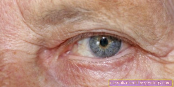
In most cases, neurocutaneous melanocytosis is asymptomatic, that is, in the case without the involvement of neurological structures. If there is neurological involvement, nevus melanocytes also accumulate in the central nervous system. Such cases lead to clinical symptoms including headache, seizures, vomiting, visual disturbances, movement disorders and paralysis. With neurological involvement, there is a risk of developing a tumor of the connective tissue in the meninges (leptomeningeal melanoma) or of malformations of the brain (e.g. internal hydrocephalus).
Treatment of neurocutaneous melanosis
A regular annual check-up of moles by a dermatologist is important, as they pose a high risk of degeneration. This means that the nevi can develop into melanomas (skin cancer).
During the first weeks of life of an infant suffering from neurocutaneous melanocytosis, the large-area nevi can be ground off (dermabrasion). However, because there are numerous nevi, this is not entirely possible. Conspicuous nevi must therefore be observed or, if in doubt, cut out.
Read more about this: Remove the birthmark
If neurological symptoms such as internal hydrocephalus occur, neurosurgical surgery must be considered. Those affected must also be neurologically monitored for life using MRI and computed tomography (CT).
forecast
Patients with asymptomatic neurocutaneous melanocytosis have a normal life expectancy.
In contrast, a disease with neurological symptoms has an unfavorable prognosis. The majority of those affected die within the first three years after the symptoms develop, as the risk of developing a tumor in the meninges is high.
In addition, other malformations of the brain often occur, with many patients developing internal hydrocephalus ("water head"). This is an enlargement of the fluid-filled cavities (ventricles) in the brain. The cerebrospinal fluid (cerebrospinal fluid) usually drains from the brain into the spinal cord. In neurocutaneous melanocytosis, birthmarks form in the area of the drainage, which enlarge over time and thus impede the drainage of the CSF from the brain. As a result, intracranial pressure rises and can seriously damage the brain.
Read more about this: Water head