Kidney stones
Synonyms in a broader sense
Urolithiasis, nephrolithiasis, urinary stones, kidney stones
English: renal calculus

Definition of kidney stones
Kidney stones (Urolithiasis) are defined as urinary stones in the kidney and the urinary tract (Urinary stones). These kidney stones are caused by disturbances in the chemical balance of the urine. ly it is about crystalline structures. The size and location of the stones as well as any sequelae determine the symptoms that arise (Symptoms).
frequency
Every year 1% of men and 0.5% of women in Germany develop kidney stones. The probability of suffering from it at some point in your life is approx. 4%, so kidney stones / urinary stones are more common than z. B. Diabetes mellitus (3%). Adults between the ages of 20 and 50 are most commonly affected.
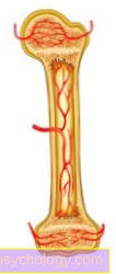
Figure kidney stones

Kidney stones - nephrolites
(Urinary stone formation in the kidney)
- Right kidney -
Ren dexter - Ureter -
Ureter - Urinary bladder -
Vesica urinaria - Female urethra -
Urethra feminine - Prostate gland - prostate
- Male urethra -
Urethra masculina - Kidney bay with filling fat -
Renal sinus - Renal pelvis - Pelvis renalis
- Calyx
(Part of the renal pelvis) -
Calix renalis - Renal medulla - Medulla renalis
- Renal cortex - Renal cortex
(A to E - kidney stones)
A. - Kidney stone on entry
into the ureter
B - renal pelvic stone
C - kidney stone in the kidney bay
D - calyx neck stone
E - Chalice
F - ureteral stone
G - bladder stone
H - urethral stone
(left female pelvis,
right male)
I - prostate stones
You can find an overview of all Dr-Gumpert images at: medical illustrations
Causes of Kidney Stones
The formation of kidney stones is very complex and only partially known to this day.
1. Prerenal causes of kidney stones (i.e. the cause lies in front of the kidney through an increased attack of urinary stone-forming substances, especially calcium and phosphate)
- Vitamin D overdose (Rare)
- Immobilization (immobility) as a result of an illness: bone remodeling or degradation can lead to a disruption of the calcium and phosphate balance (= increased excretion in the urine)
- Hyperparathyroidism: overactive parathyroid glands (due to the increased production of hormones, there is increased calcium and phosphate excretion in the urine); responsible for 5 - 10% of all calcium-containing urinary stones!
2. Renal causes of kidney stones (the cause is in the kidney itself)
- Renal tubular acidosis: Defect of the kidney tubules (insufficiently acidic urine can not be formed - pH value never below 5.8)
- Hypercalciuria: calcium excretion of more than 8 mmol / d (e.g. due to reduced uptake in the kidneys or increased uptake via the intestine)
3. Postrenal causes of kidney stones (the cause is in the lower urinary tract)
- Urinary flow disorders
- Urinary tract infection / cystitis
Read more on this topic at: Causes of Kidney Stones
Kidney stones and stress
stress and severe psychological stress can be triggers for kidney stones. The formation of stones in the urinary tract is based on an interplay of various factors, such as diet, exercise, water balance, age and many more. Stress is only one component that can have a negative impact and should be taken into account during treatment or prevention.
Kidney stones caused by alcohol
Alcohol, especially beer, leads to an increase in uric acid in the blood, a breakdown product of so-called purines. Since uric acid stones make up the second largest proportion after the oxalate stones, alcohol is a major risk factor for the development of kidney stones. Alcohol also has a flushing effect, but the negative consequences outweigh the consequences.
Read more on the topic: Consequences of alcohol and kidney pain after alcohol
Symptoms / complaints
1. Symptoms of kidney stones
ly kidney stones are found in the calyx system, in which the kidney stones often cause no symptoms (so-called "silent" kidney stone). However, the kidney stone can also trigger colic (wave-like, cramp-like pain with symptom-free intervals) if it moves from the renal pelvis into the ureter and passes through several constrictions.
Depending on where the kidney stone is, the pain radiates in a typical way. With kidney stones, the lumbar region is mainly affected ("Lower back pain"). Kidney stones that have already reached the urinary bladder lead to a painful and frequent urge to urinate, with the pain radiating to the penis and testicles or the clitoris and labia.
Read more on this topic: How can I relieve kidney pain?
Nausea and vomiting occur more frequently with colic. The stomach is bloated. It can be accompanied by reflexes in intestinal paralysis (Ileus) come, the heart beats slower (Bradycardia). Fever is only found if there is a simultaneous urinary tract infection (cystitis, pelvic inflammation)
Those affected are restless and writhed in pain, which is repeatedly interrupted by pain-free periods.
2. Symptoms of bladder stones
The bladder stones are usually caused by a urine outflow problem, e.g. B. Enlarged prostate (prostatic hyperplasia). In rare cases, they can grow up to the size of a hen's egg. Since they usually do not obstruct the exit of the bladder, they cause few symptoms: frequent urination (pollakiuria) and occasionally blood in the urine (hematuria). Furthermore, lower abdominal pain, interrupted urination and the urge to urinate that cannot be suppressed can occur.
diagnosis
Patients with:
- already affected family members
- inflammatory bowel disease (e.g. Crohn's disease, ulcerative colitis)
- osteoporosis
- Small bowel surgery
- History of kidney stones
When diagnosing kidney stones, the urine is checked for red blood cells and bacteria using test strips and sediment analysis (you can look at the solid components). In this way, blood leakage through the urine and a urinary tract infection are examined. The pH value should also be checked several times, because deviations in the case of corresponding complaints may indicate kidney stones / urinary stones.
In blood (laboratory) calcium, phosphate, chloride, creatinine and uric acid (indication of urinary stone-forming substances) are checked.
If there is then a suspicion of kidney stones, this must be confirmed by an ultrasound and X-ray examination. If the kidney stone cannot be seen on the X-ray, it must be taken into account that not all stones cast a shadow that is visible in the X-ray or that other causes can be behind a urinary outflow disorder (see above). In rare cases, vascular occlusion can also cause colic-like pain.
A Urogram May only be carried out in the colic-free interval, as otherwise there is a risk of a ureter rupture. Here, kidney stones are diagnosed iodinated contrast agent introduced into the vein and then excreted by the kidneys. After that, after 7 and 15 minutes X-rays made on which the kidneys, renal pelvis, ureters and bladder become visible and possibly non-radiopaque stones unmask themselves by the contrast agent washing them around.
You should also clarify whether the cause of the stone formation is to be found in the patient's metabolism (enzyme defects, etc.). For this purpose, eating habits, drinking behavior and ingested Medication queried.
The diagnosis is expanded for children and patients with recurring urinary stones. It'll be twice 24h urine collected and based on calcium, magnesium, PH value, Uric acid, creatinine, cystine, oxalate, citrate and phosphate were examined. Deviating values indicate metabolic disorders already described above.
The choice of Therapy of kidney stones depends on the location of the stone in the urinary system, its size and kidney function.
Differential Diagnoses / Alternative Causes
- Gallstones: You can Biliary colic causing pain in the shoulder and mid-abdomen.
- Appendicitis (appendicitis): There is no typical colic, but rather a permanent pain with tenderness in certain areas.
- In women it is also pinched off Ovarian cysts or one Ectopic pregnancy to think.
Removal of kidney stones
About 4 in 5 kidney stones are eliminated from the body without having to be removed. Regular checks are sufficient in such cases. Even if pain is in the form of a so-called renal colic may have occurred through conservative treatment in the form of Fight pain, Heat application, physical exercise and adequate hydration a spontaneous stone loss be obtained with the urine.
Through regular checks using Ultrasonic or roentgen the urologist should check whether the measures are successful. There are various ways of removing larger stones or if they do not come off spontaneously and cause pain. Urinary stones, the cause of a Urinary tract infection or one Urinary flow disorder should also be removed regardless of their size.
Removal of kidney stones with the help of a laser
Kidney and other urinary stones can be targeted with a Laser probewhich is pushed into the body through an endoscope to the stone, are crushed. The endoscope can be advanced through the natural body openings, via the urethra and into the bladder. Another option is access via a small incision below the costal arch and a thin tube that is pushed into the renal pelvis. The stone fragments resulting from the cutting with the laser are removed via the endoscope. Injuries caused by the laser are very rare among experienced surgeons.
Removal of kidney stones with the help of an operation
Kidney stones from a size of about 1 cm are usually removed by the urologist via an endoscopic operation general anesthetic carried out. The procedure is called minimally invasive percutaneous nephrolithoapaxy or short Mini PNL designated. A thin tube is placed over a small skin incision (about 1 cm) on the side below the ribs under visual control, using ultrasound, up to the renal pelvis.
An endoscope is advanced through this so-called puncture channel. In addition to a camera, this is equipped with a laser probe. So can bottom view a targeted crushing of the kidney stone can be carried out. The fragments are immediately flushed out through the endoscope. An inpatient stay of three to five days is usually necessary for the procedure. Complications are rare.
In contrast to the minimally invasive operation described, the open surgery only performed in exceptional cases for kidney stones, for example if the entire renal cavity is filled with a huge stone or other stone removal methods are unsuccessful.
Removal of kidney stones with a sling
To remove kidney stones with a snare, an endoscope about 3 to 4mm wide is inserted through the urethra. In addition to a camera, this has a Working channelover which the loop is pushed into the bladder and then over the ureter to the point where the kidney stone is located. This is covered with the noose and ultimately pulled out of the body. The procedure is performed under general anesthesia. An inpatient stay is necessary for this.
Smash kidney stones
You can use kidney stones mechanical pressure waves can be deliberately shattered from the outside into small fragments, which are then excreted in the urine. The procedure is called extracorporeal shock wave lithotripise (ESWL) designated. The waves spread through the skin and develop their effect on the kidney stone. The method is painless, however, it can be due to damage to kidney tissue temporarily to excrete blood in the urine come. Many patients complain about that Noise pollutionl found very uncomfortable during the examination. One in three patients treated has extremely painful symptoms after stone fragmentation Colic (severe cramping, labor-like pain) caused by the loosened stone fragments.
This method is preferred for smaller, individual stones. The success rate is around 80%. However, if the location of the stone is unclear, the chances of success are lower. Advantages over other methods are the lack of invasiveness, which means it must no skin incision and anesthesia is not required. That's why I have to no inpatient stay but it can be a outpatient treatment be performed.
Home remedies for kidney stones
The most important recommendation for prevention and treatment of kidney stones is much to drink. Heat pillows, bottles or envelopes in the flank area can be helpful. If the pain allows, the stone can be loosened by climbing stairs or hopping. There are also several natural ways to increase the flow of urine with the aim of excreting the kidneys or ureteral stones: Beside Kidneys- and Bubble tea Cranberry juice from the pharmacy is recommended.
Also effective is a tea made from dried dandelion. To do this, pour two teaspoons of boiling water over it and let it steep for five minutes. For cramp-like pain (colic) can Broken herb Provide relief. This time, cold tap water is poured over a teaspoon and left to stand for 30 minutes. Before drinking, this brew is boiled for three minutes. Occasionally will Beer enjoyment recommended for urinary stones due to its flushing effect. Due to the purines it contains, however, it can increase the risk of urinary stone formation and should therefore tend to be avoided.
How can you prevent kidney stones?
To prevent kidney stones from occurring for the first time or again, it is particularly important to always much to drink, preferably two to three liters of water or herbal tea daily. Black tea or coffee, on the other hand, can increase the risk of kidney stones due to the oxalate they contain and should therefore be avoided. Also is regular exercise For example, walking is helpful to prevent kidney stones from forming and to enable smaller stones to pass before they become larger and cause pain. In the diet, animal fats should make up a small proportion. Due to the different possible components of kidney stones, certain foods should be avoided after suffering from illness in order to prevent kidney stones from recurring. Appropriate advice should be given by the doctor. Since being overweight is also a risk factor for the formation of kidney stones, it is advisable to reduce this if necessary.
Figure kidney

- Renal cortex - Renal cortex
- Renal medulla (formed by the
Kidney pyramids) -
Medulla renalis - Kidney bay (with filling fat) -
Renal sinus - Calyx - Calix renalis
- Renal pelvis - Pelvis renalis
- Ureter - Ureter
- Fiber capsule - Capsula fibrosa
- Kidney column - Columna renalis
- Renal artery - A. renalis
- Renal vein - V. renalis
- Renal papilla
(Tip of the kidney pyramid) -
Renal papilla - Adrenal gland -
Glandula suprarenalis - Fat capsule - Capsula adiposa
You can find an overview of all Dr-Gumpert images at: medical illustrations