Pigment spots
Introduction / General
The pigment spots (Syn. Pigmentary nevus, melanocyte nevus, melanocytic nevus) is an initially benign malformation of the skin, which arises from pigment-forming melanocytes or related cells.
For this reason, the pigment spots are often colored brown. There are numerous subspecies of benign pigment spots, which in some cases degenerate and thus become malignant (malignant) can be.
Pigment disorders in the face and pigment disorders in the neck are particularly common.

Remove pigment spots
Most of them Pigment disorders are completely harmless and represent at most a cosmetic problem. For this reason, a Removing the pigment spots rarely necessary. However, if you decide to treat the pigment spots, there are various methods to choose from.
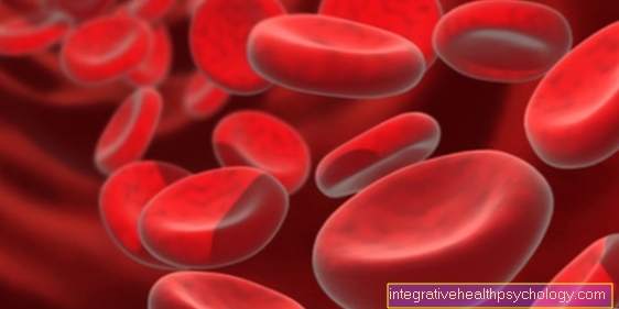
It is very effective Laser treatment, in which pigment accumulations are shattered by the bundled energy of the laser and the remains are subsequently removed from white blood cells be dismantled. Another option is that Cold therapy (Cryopeeling) with liquid nitrogen or a treatment with acids. These ensure that the upper ones die off Layers of skinso that they can be removed together with the melanin it contains. However, the sensitive skin tends to form new pigment spots in the subsequent period and should be protected from direct sunlight. A widespread form of treatment for pigment spots is also the use of bleaching creams based on Rucinol, Hydroquinone or Kojic acidwhich, however, are potentially hazardous to health and often do not show sufficient effect.
In addition to cosmetic aspects, a degeneration of the pigment spots can also be a reason to have them removed. Anomalies are usually difficult to recognize for the layperson. Nevertheless, it is advisable to keep an eye on pigment disorders and, above all, to pay attention to changes in the pigment spots.
Laser pigment spots
Laser procedures are a very effective method of treating Pigment disorders, in which pigment accumulations are shattered by the bundled energy of the laser and the remains are then broken down by immune cells.
There are different types of lasers such as ruby-, Erbium-, KTP- or Fraxel laser to disposal. They differ in terms of their wavelength and depth of penetration.
Several sessions are usually necessary to completely remove the pigment spots. If a lightening of the pigment disorder is sufficient, a single laser may be sufficient. It is best to do this kind of treatment in autumn or winter, as the UV radiation is lowest during these times of the year. This is noteworthy because the skin is very sensitive after a laser treatment. In addition, it must be covered with sunscreen every day for at least eight weeks after the laser. The laser treatment always carries the risk of scarring and should therefore be used carefully.
The cost of a laser treatment for pigment spots is around 100 euros per session.
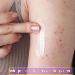
A cream used to treat pigment spots
Before removing the pigment spots with a laser or lightening them with cold or acid treatment, simpler means such as creams can be used. Many, especially prescription only, Creams are based on the effect of the bleaching agents contained. These interrupt the formation of melanin in the melanocytes. A very common bleach is among others Hydroquinone. However, since this is suspected of being carcinogenic (carcinogenic), creams containing hydroquinone should only be used for a maximum of 3 months. Other common bleaches are Rucinol and Kojic acid.
A treatment with creams containing bleach is usually only successful after about two months; light pigment spots can be significantly lightened after about four weeks. To avoid undesirable side effects when using creams containing bleach, a doctor should always be consulted before using them.
Please also read our topic: Bleach for the skin
Cell types
The pigment spots can arise from different cell types and accordingly have different characteristics.
The melanocytes are the body's melanin-producing cells and lead to the formation of brown pigment spots.
Depending on the type of cell, these pigment spots are called melanocytic nevi designated. According to their location in the skin layer, the Pigment spots further divided into:
- epidermal melanocytic nevi and
- dermal melanocytic nevi
The nevus cells are closely related to the Melanocytesbut have none Dendrites. They lie in the skin in the form of spherical to spindle-shaped cells that are arranged in nests. In addition, they cannot release their pigment to the surrounding skin cells.
In addition to the benign (benign) There are also numerous atypical cells that are malignant (malignant) can degenerate. These malignant cells can arise from melanocytes as well as from nevus cells. These cells then lose for various reasons (UV light, Genetics, wrong repair mechanisms, ...) their normal shape and growth rate and degenerate.
Melanocytic nevi
These pigment spots arise from real melanocytes and are divided into epidermal and dermal melanocytic nevi.
To the epidermal pigment spots (pigment disorder) include the following types of nevus:
1. Freckles (Ephelids): These are small yellowish and brownish spots on the skin, which occur especially in people with a fair complexion and red or blond hair.
Freckles are pigment deposits that are mainly caused by sunlight. In some people, the freckles fade again in winter.
That of the Melanocytes The melanin produced is stored in the surrounding keratinocytes and thus leads to the brown coloration of the skin. In contrast to the Moles there is no local increase in melanocytes. In most cases, freckles are a norm variant, which is caused by an innate genetic variation in the Melanocortin-1 receptor arises. In the case of the NAME syndrome, the freckles are a symptom of a systemic disease, which is accompanied by other Skin changes goes hand in hand.
2. Lenticular nevus: This is a benign, sharply edged one brownish mole. Of the "Lentigo simplex“Refers to the normal mole, which occurs in children through the influence of sunlight. It is flat, brown, round or oval and usually smaller than 5mm in diameter.
Histologically, there are no nests of nevus cells, as in a junction nevus, but only an increased number of melanocytes. For this reason, the lentigo simplex cannot be black Skin cancer (malignant melanoma) arise.
The lentigo solaris (Age spots) are also mainly caused by long-term sun exposure. The age spots multiply increasingly in late adulthood. It is a flat, brown lesion at the level of the skin with a blotchy or star-shaped outline that is always sharply defined. Usually the stain is brown and irregularly pigmented.
The age spots mainly affect light skin types. While they are not cancerous, they can be mistaken for black skin cancer. Despite everything, they are an expression of sun damage and for this reason only occur on areas of skin that are exposed to the sun.
3. nevus pigmentosus (Café-au-lait spots): These are always benign, light to dark brown, uniform ones Moles. You can choose between 2mm up to 20cm be large and, unlike other pigment spots, never are sublime or knotty. Because of this, the Café-au-lait spots no disease value and can be found at 10- 20% of the normal population. If there are more than 6 spots, each larger than 15 mm in adulthood, or larger than 5 mm in childhood, as well as other symptoms, the diagnosis is one Type I neurofibromatosis probably.
4. Spilus nevus („Lapwing egg nevus“): This one Pigment spot is characterized by a large, evenly brown-colored patch of skin, which also has numerous small dark brown spots. The overall diameter is often larger than 15cm, the small brown spots often have a diameter of 2-3mm on.
On average, this form of pigmentation occurs in 3 out of 100 fair-skinned adults. The small dark brown spots can be atypical, the development of black skin cancer (malignant melanoma) is very rare, however.
5. Becker nevus: This type of pigment spot mainly affects young men. Typically, a palm-sized, sharp and jagged border forms in the shoulder area hyperpigmented Spot. This arises exclusively due to increased melanin formation. The skin change should be completed within a year and then no longer change.
As a rule, the Becker nevus only rarely turns pale afterwards. It has no malignant potential, so that it can be removed using laser therapy for purely cosmetic reasons.
To the dermal melanocytic nevi belong to the following subgroups:
1. Mongolian spot (Rump spot sacral spot): This spot is usually irregular and bluish. It is located on the back, buttocks or sacrum in newborns and is harmless.
It is a remnant of the embryonic development and fades or usually disappears afterwards 4 to 8 years or at the latest by puberty. Overall, the Mongolian spot is much more common among Asians among Black Africans. It is seldom found in fair-skinned and blonde children.
2. Fusco-coeruleus nevus: It is a dark bluish to brownish one Pigment disordercaused by the ectopic accumulation of melanocytes in the deep Dermis (Dermis) arises. Because of its two different locations, it is called Nevus ota and Ito nevus designated. Of the Nevus ota mostly affects the supply area of the first and second trigeminal branch and is therefore located on the forehead, eye area, cheek and palate.
He can do that too Conjunctiva, Sclera and the eardrum to include. The Ito nevus is located in the shoulder area. In exceptional cases, this pigment spot can become malignant. Since the nevus ota in particular can be very disfiguring, it is possible to treat it with laser therapy.
3. Coerule nevus (blue nevus, dermal melanocytoma): It is characterized by a dark blue to gray-black color, sharply defined and benign.
The unusual color is due to the accumulation of melanocytes in the deeper layers of the skin. It is assumed that the melanocytes accumulate ectopically in the deeper layers in the course of development. The blue nevus is most common on the back of the hand and the back of the forearm. However, it can appear anywhere and is usually harmless. The development of a malignant melanoma is very rare, so that the removal can take place for purely cosmetic reasons.
Nevus cells (nevus cell nevus)

These cells have none Dendrites and cannot deliver their pigment to neighboring cells. Typically, the nevus cell nevus develops during childhood and in some cases regresses completely.
1. Junction nevus: In this first stage, the nevus grows exactly on the border between epidermis and Dermis (Dermis). This zone is called the junction zone. It is sharply demarcated, punctiform, brown or black. These first junction nevi arise in childhood.
2. Compound nevus: This is the second stage of the Nevus cell nevus- development. The nevus moves into the depths of the dermis and thus expands into both layers of the skin. The surface of the pigment spot can be fissured. This will make the Mole overall thicker and may have knotty parts. During this phase, the pigmentation often becomes more irregular and lighter.
3. Dermal nevus: This is the final stage in the development of a nevus cell nevus. Often it takes on a large, round, hemispherical shape. The nevus cells have completely penetrated the dermis and the nevus has usually lost its brown pigment and is with Hair occupied.
4. Congenital nevus: These pigment spots are already at the birth available (connatal) and are light to dark brown in color and often have a knotty to cobblestone-like surface. This Pigment spots can from 1.5cm to over 40cm be great. In this case, they are called giant nevi, which are most commonly found on the stomach or back.
In addition, these giant nevi can have bristly hair, which is why they are known as animal fur nevus. The greater the size of the congenital nevus, the greater the risk of developing malignant melanoma, which is why giant cell nevi should be removed in the first year of life.
5. Halonevus: These pigment spots are characterized by a white, pigment-free ring. It occurs mainly in childhood through to young adulthood. It is believed that autoimmunological processes cause the destruction of melanin and melanocytes and thus promote the development of white spots. As a rule, the halo nevus disappears after a while and is harmless.
6. Pointed nevus: These nodular, benign pigment spots occur mainly in children and adolescents. It grows quickly and is reddish to brown in color. It is often hemispherical, coarse and hairless. In diameter it is usually smaller than 1 cm. Usually this type of nevus does not resolve on its own. However, they are usually benign, but they can look similar to malignant melanoma and are therefore easy to confuse.
7. Dysplastic nevus: The dysplastic nevi occur in approx. 5% of people before.
They show a more restless image on the skin than normal nevus cell nevi. They are often pigmented differently and their borders are often blurred with frayed edges.
They are usually larger than 5mm and some of them can have raised parts. Moving from a dysplastic nevus to a malignant melanoma can take months or years, with the risk of developing one malignant melanoma increases in the presence of dyplastic nevi of 0,8% on 18%.
For this reason, dysplastic nevi that change or appear conspicuous should be completely removed.
Pigment spots on the face
As Pigment spots (Hyperpigmentation) is the term used to describe the brown discoloration of the skin caused by the activation of Melanocytes are conditional.
This activation takes place primarily through the UV radiation contained in sunlight. For this reason, pigment spots are very often found on the face, shoulders and hands.
Pigment spots can be in the form of Freckles (Ephecids) or age spots (Lentigo solaris) occur and take on various shades of brown, reddish or ocher. A very common special form of pigment disorders is the Café-au-lait spot , which owes its name to the light to dark brown, very even pigmentation. However, it is not only found specifically in the facial area.
The so-called Pregnancy mask (Chloasma) is hormonal. In many pregnant women, the nipples temporarily darken during pregnancy and the typical brown line from the navel to the pubic bone (Linea nigra) forms.
There can also be sharp, irregularly defined pigment spots on the face. They are predominantly found on forehead, Temples, Cheeks and chin and are often very symmetrical.
Since the pregnancy mask usually regresses by itself after pregnancy, bleaching agents are not recommended. Instead, cosmetics can be used if the pigment spots are very annoying. In order to at least partially prevent pregnancy masks, it is advisable to use sunscreen and not expose yourself to intense sunlight.
Some drugs have the property of increasing the skin's sensitivity to light and can thus also contribute to the formation of pigment spots. These include certain antibiotics (especially tetracyclines such as doxycycline, which is often used in urinary tract or gastrointestinal infections), chemotherapeutic agents and also St. John's wort preparations.
If pigment disorders on the face are perceived as very annoying or if pigment spots are suspected of degenerating, these can be lightened or removed, just like on other parts of the body, using various treatment methods such as lasers or bleaching creams.
Forecast and summary
Due to the large number of pigment spots and the potentially malignant degeneration should Birthmarks be examined at regular intervals by a dermatologist.
Freckles, Café-au-lait spots and small spots Lentigenes consist of normal melanocytes and are not at risk of developing a malignant melanoma. The development of a malignant melanoma, however, is directly related to the number of existing nevus cell nevi.
Dysplastic nevi can arise directly from nevus cell nevi and increase the risk of melanoma by 100 times. Nevi that are already present at birth (congenital nevi) promote the development of malignant melanoma.


.jpg)