Plica Syndrome
definition
Under a Plica Syndrome if you grasp one Symptom complex together, the most of all Pain and Movement impairments exists in the affected organ system. The cause of a plica syndrome lies in one Skin foldthat has not receded as it should in the course of life.

Cause / forms
A Plica is a physiological skin foldthat exist in various organ systems of the human body. The task consists of one Formation of reserves on the skin, in the mechanically stressed regions of the body is necessary. This fold of skin is largest at birth and regresses over the course of life. In some cases it is no longer detectable in adulthood, in some cases remains of this skin fold still remain.
The so-called Plica syndrome it comes to one incomplete regression this fold of skin.
In the knee joint area there is also a plica that extends from the inside of the knee joint in a central direction. There is only limited space available in the knee joint due to the anatomical conditions. The not completely receded plica fits into the knee joint, but can lead to discomfort. These complaints are caused on the one hand by friction between the cartilage and plica with every movement, on the other hand pain can result from pinching the plica in the joint space.
In the case of friction between the plica and cartilage of the joint surface, the patient does not initially notice any discomfort, as the cartilage still has a protective effect. With increasing friction, however, the cartilage is worn down and the bone in the area of the joint surface is exposed. As soon as bone is exposed and the skin fold rubs against the bone, discomfort in the sense of pain occurs. The pain can be triggered especially when moving.
At the beginning of a plica syndrome, when the cartilage is still partially preserved, complaints in the knee joint only occur after heavy loads. However, as soon as it is an advanced plica syndrome and bone is increasingly exposed, on which the plica rubs, complaints increasingly arise even at rest. In addition to the pain, which can occur either depending on movement or at rest, the pain-plagued patient will also adopt a relieving posture in order to alleviate the corresponding symptoms. If carried out for a long time, this relieving posture also leads to poor posture, which in turn can lead to complaints.
Please also read: Painful fold of mucous membrane in the knee
Appointment with a knee specialist?
I would be happy to advise you!
Who am I?
My name is I am a specialist in orthopedics and the founder of .
Various television programs and print media report regularly about my work. On HR television you can see me every 6 weeks live on "Hallo Hessen".
But now enough is indicated ;-)
The knee joint is one of the joints with the greatest stress.
Therefore, the treatment of the knee joint (e.g. meniscus tear, cartilage damage, cruciate ligament damage, runner's knee, etc.) requires a lot of experience.
I treat a wide variety of knee diseases in a conservative way.
The aim of any treatment is treatment without surgery.
Which therapy achieves the best results in the long term can only be determined after looking at all of the information (Examination, X-ray, ultrasound, MRI, etc.) be assessed.
You can find me in:
- - your orthopedic surgeon
14
Directly to the online appointment arrangement
Unfortunately, it is currently only possible to make an appointment with private health insurers. I hope for your understanding!
Further information about myself can be found at
Symptoms
At the onset of the syndrome, severe physical exertion, such as climbing stairs or hiking in the mountains, causes symptoms.
With advanced syndrome and increasingly exposed bones, discomfort can also arise at rest.
The entrapment immediately leads to acute complaints, which can be very severe. In this case, the patient feels pain even with light strain in the sense of knee flexion.
Find out more about: Acute knee pain - that may be behind it
Diagnosis

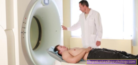
The diagnosis one Plica Syndrome is difficult. Besides the physical exam, in which above all diseases of the meniscus and the ligamentous apparatus are excluded, which can lead to similar complaints, should always be one imaging diagnostics be performed. Because a normal X-ray examination of the knee mainly shows bony changes and less the soft tissues, a simple X-ray is not the method of choice.
Rather, one comes Magnetic resonance imaging (MRI) of the knee for use. There the spatial conditions in the knee joint area can be displayed and it can be seen whether a Plica is available. If this is the case, it can also be assessed whether the plica has become trapped in the joint space of the knee joint.
Also comes as a diagnostic tool Knee arthroscopy for use in which a camera can be brought into the knee joint in addition to instruments through small skin incisions above the knee. The advantage of this method is that on the one hand Real images of Knee joint can be transmitted to the examiner and, on the other hand, the patient's knee joint can also be moved during the examination. Sometimes it is the case that an existing plica can become trapped in the knee joint during flexion and slip back into its normal position during extension. This could be done with the Arthroscopy recognize well.
Often times, the diagnosis of plica syndrome is rather one Differential diagnosis if no illness belonging to the complaints can be seen.
MRI for Plica Syndrome
An MRT examination is generally very helpful if the ligaments of the knee joint are to be displayed, as soft-tissue body structures are also clearly depicted. However, since the plica is often entrenched in the joint in a rather unfavorable way, it cannot always be made visible via an MRI examination. In these cases, the diagnosis can either be made through a pioneering physical examination or, ultimately, only during the arthroscopy itself.
Read more on the subject at: MRI of the knee joint
therapy
Often one is enough conservative therapy out. This is especially true for plica syndromes, where still enough space in the joint space exists and it too no cartilage degeneration has come.
This is definitely one of the conservative treatments Reduce stressful movements. Excessive sport should be reduced or completely avoided, movements that place particularly high stress on the knee joint (climbing stairs, mountain hiking) should be reduced. The knee should regularly chilled become. Also can anti-inflammatory drugs, how Ibuprofen or Diclofenac are used. If these measures are not sufficient, a arthroscopic removal of the Plica is an operational measure that can be carried out. Ideally, the plica can be ablated and removed from the joint space using the instruments introduced during the diagnostic treatment. Another conservative measure is one physiotherapy treatmentwhich should be connected in any case, even after a surgical procedure. In this physiotherapy, the Musclessurrounding the knee joint, trained and thus that Knee joint spared. Physiotherapy should be carried out consistently and regularly over several weeks.
Who needs an operation?
First of all, it can be stated that not every plica necessarily requires therapy. It is estimated that there is such a synovial fold in about every second knee joint. But not everyone has complaints because of it. The plica only becomes disturbing when the knee joint is subjected to heavy loads, e.g. Squatting down or cycling frequently, causing joint pain. In this case, therapy to alleviate the symptoms should be initiated.
An operation is only considered if conservative therapy methods have failed and the pain persists or the knee is severely inflamed. Conservative measures include protection and cooling of the inflamed joint, physiotherapy, a supply of suitable insoles for shoes, anti-inflammatory drugs or pain medicine or joint injections with the anti-inflammatory cortisone.
Even if the non-surgical therapies failed beforehand, there is an extremely high probability of recovery from the operation. However, it must be taken into account that pain may still be present after the operation if cartilage damage has already occurred due to the plica syndrome. These are not remedied by the minimally invasive surgery. In addition, if the patient is physically active, it is more likely to opt for the operation, since with constant sporting stress on the knee joint, it is not to be expected that in the long term, freedom from symptoms can be achieved through conservative measures.
Course of an operation
The plica syndrome is operated arthroscopically. This means that the knee is not opened completely through a long skin incision, but only a camera and a surgical tool are introduced into the joint in the working channel through two much smaller, lateral incisions. Controlled by the camera, the interfering synovial membrane (plica) can then be removed via the working channel. The procedure usually takes no longer than 15 minutes and is usually performed on an outpatient basis with local anesthesia.
Follow-up treatment of the operation
After the operation, drains usually remain in the operating area for about two days. In addition, walking aids are required for the first time, as the knee must not be fully loaded. Anti-inflammatory drugs are also prescribed for as long as signs of inflammation can still be seen. If necessary, physiotherapy should be used in the course of the operation to strengthen the thigh muscles that span the knee. Electrotherapy can also be used to stimulate the muscle. When doing sport, it is important to ensure that uniform movements, e.g. Cycling are possible again as soon as the knee can be bent enough. Sports with many start-stop movements, such as Tennis or soccer, on the other hand, should be avoided until the knee is free of inflammation and the knee has healed.
How long do you need crutches?
How long after the operation you have to use crutches depends on your recovery. In general, the walking aids should be used as long as the joint is still irritated. It may appear that it has healed after two to three days, but it can also take two to three weeks before the crutches can be dispensed with. In general, the extensor muscles of the thigh should be trained immediately after the operation. Complete relief would be counterproductive and prolong the healing process. Overloading should of course also be avoided.
How long are you on sick leave?
How long you will be unable to work after the operation depends on various factors. On the one hand, a generally good overall physical constitution of the patient leads to faster healing. The cooperation of the patient also plays an important role. If the necessary muscle building exercises are not carried out after the operation, this will have a negative impact on recovery and will prolong it. Once the knee has healed completely, no damage remains and full weight bearing is possible again. As a rule, you can work again after about one to four weeks. Sport is only fully possible again after about four to six weeks.
Physiotherapy after surgery
Physiotherapeutic treatment should be started immediately after the operation. The aim of the treatment is to strengthen the muscles that surround the knee joint so that the joint becomes more resilient. The exercises can primarily be performed with the help of your own body weight or with the help of training bands. It is also important to train the core muscles, as these contribute to the stability of the leg.
In the beginning, walking training can consist of slow uphill walks on the treadmill. During the course, jumping training should be introduced, which prepares for jogging in the last step. Because jogging requires a certain ability to jump, as both feet briefly leave the ground with every step.
It should also be checked whether there is a muscular imbalance between the extensor and flexor muscles of the knee. In this case, this should be remedied by specifically strengthening the weaker muscles in order to reduce the pressure on the kneecap. An imbalance between the outward and inward pulling muscles of the thigh also has a negative effect on the plica syndrome, as the kneecap is pulled out of the midline and thus incorrectly loaded. Regular stretching of the knee joint muscles can also be helpful.
Summary
Under a Plica syndrome one understands you Symptom complexin which it has not regressed Skin fold in the knee joint to Entrapment or Frictions can come. Due to the limited space in the knee area, the joint cartilage, which is becoming increasingly thinner, will soon rub. In this case there are initially no complaints. When the articular cartilage has released bone in certain places, skin folds come into direct contact with the bone, which usually leads to Relief postures and Movement impairments. Especially movements that put stress on the knee joint, such as Climbing stairs or hiking in the mountains can make the symptoms worse. Diagnosing plica syndrome is not easy. An MRI examination can prove the presence of a plica in the knee joint and show the available space. Often, however, entrapment cannot be shown.
The safest method a plica syndrome is proven Joint arthroscopy. Instruments are inserted into the knee joint through small incisions in the skin. The knee can also be moved during the examination and it can be assessed whether there is entrapment when the knee is flexed. Ideally, in same session a Plica removed become. This measure represents the surgical treatment of a plica syndrome. Numerous conservative methods, such as the Protection, Pain management or physical therapy but should be tried beforehand.